
No products in the cart.: $0.00

On Sale!



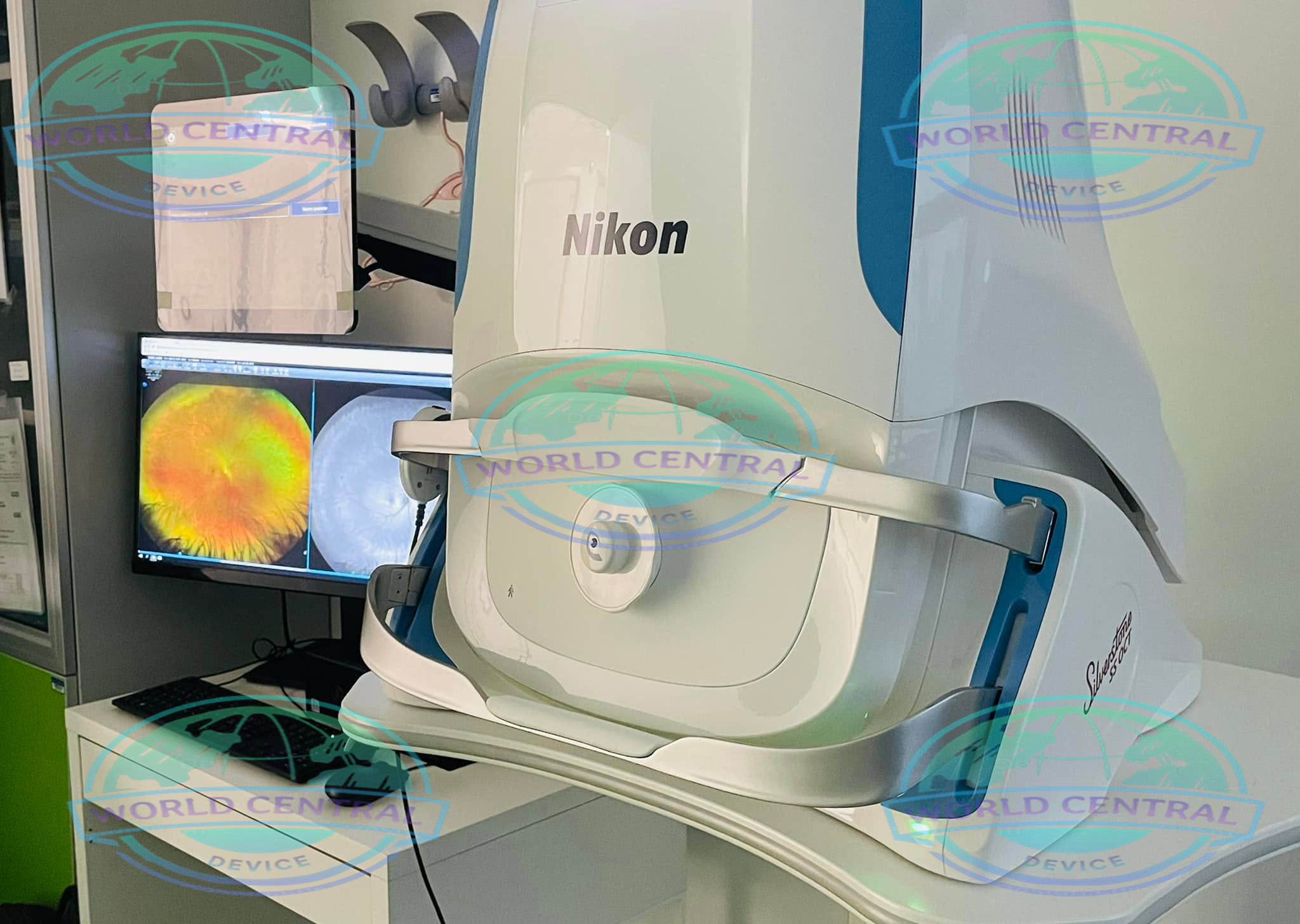
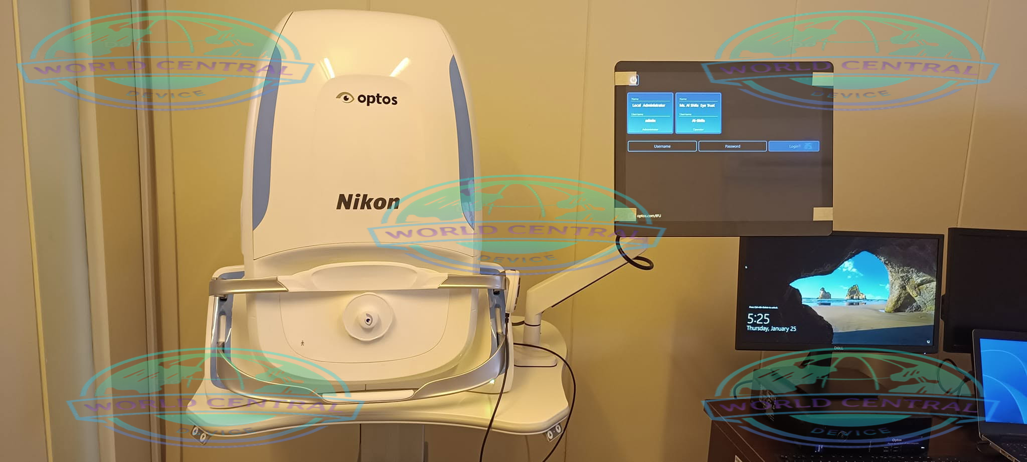

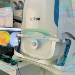
Optos Silverstone SS OCT Retinal Imaging System
The Optos Silverstone SS OCT Retinal Imaging System combines 200° ultra-widefield Optomap retinal imaging with advanced 1050 nm Swept Source OCT, delivering high-resolution multimodal imaging and guided peripheral OCT scanning for comprehensive retinal diagnosis and disease management.
Delivery and return
Delivery and return
Shipping Information
Shipping Information
Composition and care
Composition and care
Description
Optos Silverstone SS OCT Retinal Imaging System
Advanced Ultra-Widefield Retinal Imaging with Guided Swept Source OCT
The Optos Silverstone SS OCT Retinal Imaging System is a premium ophthalmic imaging platform that combines Ultra-Widefield (UWF) retinal imaging with integrated Swept Source Optical Coherence Tomography (SS-OCT). Designed for retina specialists, ophthalmologists, hospitals, and advanced eye clinics, Silverstone enables clinicians to capture a 200° retinal image in a single shot while performing targeted OCT scans anywhere across the retina—including the far peripheral retina.
Unlike conventional OCT systems that primarily focus on the posterior pole, Silverstone introduces optomap-guided SS-OCT, allowing clinicians to investigate peripheral retinal lesions with remarkable speed and precision. This combination improves disease detection, documentation, diagnosis, treatment planning, and long-term patient monitoring.
Why Choose the Optos Silverstone SS OCT?
Modern retinal diagnostics demand more than high-resolution macular imaging. Peripheral retinal diseases often develop beyond the field of view of traditional OCT devices. Silverstone addresses this limitation by integrating ultra-widefield imaging with deep-penetration swept source OCT.
Major advantages include:
- Integrated Ultra-Widefield retinal imaging and SS-OCT
- 200° single-capture retinal image
- Guided OCT scanning anywhere on the retina
- Deep choroidal visualization using 1050 nm swept source laser
- Fast image acquisition (<0.4 seconds)
- Excellent patient comfort without routine dilation in many cases
- High-speed workflow for busy ophthalmology clinics
- Comprehensive multimodal retinal imaging
- Ideal for diagnosis and follow-up of retinal pathology
- Reduced need for multiple imaging devices
Features and Benefits
UWF with integrated swept source OCT, facilitates detailed examination of the retina-vitreous to sclera
High resolution optomap image enhances pathology detection and management from macula through the far periphery1
Fast, single-capture, 200° imaging (less than ½ second), can improve clinic flow and patient satisfaction2
UWF guided, swept source OCT, images pathology anywhere on the optomap
Automatic rescan function for fast, precise follow-up scanning
Non-mydriatic, cSLO imaging, effective through most cataracts and small (2mm) pupils
1050 nm OCT light source, provides deeper tissue penetration for clear, detailed choroidal imaging
3-in-1 Color Depth ImagingTM provides important clinical data from the retinal surface through the choroid
OptosAdvanceTM Image Management software streamlines image review and consultations
DICOM compatible software supports compliance with the Code of Federal Regulations
Ultra-Widefield 200° Imaging
Silverstone captures approximately 82% of the retina in a single image, significantly exceeding conventional fundus cameras.
Benefits include:
- Visualization of peripheral retinal pathology
- Improved diabetic retinopathy assessment
- Peripheral retinal tear detection
- Better documentation for retinal specialists
- Faster patient examinations
Guided Swept Source OCT
The integrated SS-OCT allows clinicians to place OCT scans directly over suspicious lesions identified on the Optomap image.
Supported scan modes include:
- Line Scan
- Volume Scan
- High Density Scan
Guided OCT enables:
- Peripheral lesion assessment
- Choroidal evaluation
- Vitreoretinal interface analysis
- Retinal thickness evaluation
- Lesion localization
Deep Tissue Imaging
The 1050 nm Swept Source laser provides excellent penetration through retinal pigment epithelium into the choroid.
This improves visualization of:
- Choroid
- Scleral interface
- Deep retinal pathology
- Posterior vitreous
- Pigment epithelial abnormalities
High-Speed Acquisition
Fast imaging minimizes motion artifacts and improves patient comfort.
Typical imaging time:
- Less than 0.4 seconds for Optomap capture
Multimodal Retinal Imaging
Silverstone combines multiple retinal imaging technologies into a single workstation.
Available imaging includes:
- Color imaging
- Red/Green laser imaging
- Autofluorescence
- Fluorescein angiography
- Indocyanine Green angiography
- Swept Source OCT
Clinical Applications
Retina Clinics
Ideal for:
- Diabetic Retinopathy
- Age-related Macular Degeneration
- Retinal Tears
- Retinal Detachment
- Peripheral Degeneration
- Epiretinal Membrane
- Macular Hole
- Central Serous Chorioretinopathy
- Retinal Vein Occlusion
Glaucoma Monitoring
Useful for:
- Optic nerve documentation
- Retinal nerve fiber evaluation
- Posterior pole analysis
Uveitis
Supports:
- Inflammatory lesion imaging
- Peripheral retinal inflammation
- Choroidal assessment
Ocular Oncology
Suitable for:
- Choroidal nevus
- Choroidal melanoma
- Peripheral retinal tumors
- Lesion documentation
Diabetic Eye Disease
Excellent for:
- Peripheral ischemia
- Neovascularization
- Macular edema
- Longitudinal monitoring
Benefits for Eye Care Professionals
Retina Specialists
- Peripheral lesion localization
- Image-guided OCT
- Enhanced treatment planning
Comprehensive Ophthalmology Clinics
- Improved workflow
- Faster diagnosis
- Reduced patient chair time
Hospitals
- High patient throughput
- Reliable documentation
- Comprehensive retinal imaging platform
Workflow Advantages
Silverstone improves clinical efficiency by integrating multiple diagnostic functions into one system.
Benefits include:
- Fewer patient transfers
- Single imaging platform
- Rapid image review
- Improved documentation
- Simplified follow-up comparisons
Specifications
| Image Modalities | optomap color and optomap plus (red and green laser): Color composite view Green laser view Red laser view optomap af (green laser): autofluorescence Swept Source Optical Coherence Tomography (OCT) |
| Resolution | optomap color: 20 μm optomap plus, af : 14 μm |
| Wavelengths | Red laser: 635 nm Green laser: 532 nm (for af ) Blue laser: 488 nm (for fa) Infra-red: 802 nm (for icg) |
| Exposure Time | Less than 0.4 second |
| Tomographic Imaging | Signal Type: Optical scattering from tissue Signal Source: Swept source OCT, Wavelength 1050 nm Optical Power: Laser safety Class-1 following IEC/en60825-1:2014(2007)Axial Resolution: <7 micron Transverse Resolution: <20 micron (in tissue) Scanners: Galavanometric with x, y pair Scan Depth: Up to 2.5mm |
| OCT Scan Characteristics | Swept Source OCT A-Scan rate up to 100k cycles/s |
| OCT Scan Types | Line Scan Volume Scan High Density Scan |
| Footprint | Width: 540 mm/22 inches Depth: 570 mm/23 inches (including chin rest) Height: 683-707 mm/27-28 inches |
| Category | Detailed Specification |
|---|---|
| Product Name | Optos Silverstone SS OCT Retinal Imaging System |
| Manufacturer | Optos |
| Device Type | Ultra-Widefield Retinal Imaging + Swept Source OCT |
| Imaging Technology | Scanning Laser Ophthalmoscopy (SLO) + Swept Source OCT |
| Field of View | Up to 200° single capture |
| Maximum Retinal Coverage | Approximately 82% of retina |
| Image Capture Time | Less than 0.4 seconds |
| OCT Technology | Swept Source OCT |
| OCT Wavelength | 1050 nm |
| OCT Signal Type | Optical Scattering |
| OCT Scan Rate | Up to 100,000 A-scans/second |
| Axial Resolution | <7 μm |
| Transverse Resolution | <20 μm (in tissue) |
| Scan Depth | Up to 2.5 mm |
| OCT Scan Types | Line Scan, Volume Scan, High Density Scan |
| OCT Guidance | Optomap Guided OCT |
| OCT Targeting | Anywhere across retina including far periphery |
| Scanner Type | Dual Galvanometric Scanner |
| Image Resolution | Optomap Plus: 14 μm; Standard Optomap: 20 μm |
| Red Laser | 635 nm |
| Green Laser | 532 nm |
| Blue Laser | 488 nm |
| Infrared Laser | 802 nm |
| OCT Light Source | Swept Source Laser |
| Laser Safety | IEC/EN60825-1 Class 1 |
| Image Capture | Non-contact |
| Pupil Dilation | Often not required |
| Patient Position | Chin Rest |
| Image Modalities | Color, RG, Red-Free, Choroidal, Green AF, Blue AF, FA, ICG, SS-OCT |
| Color Imaging | Composite Laser Color |
| Autofluorescence | Green AF and Blue AF |
| Fluorescein Angiography | Supported |
| Indocyanine Green Angiography | Supported |
| Peripheral OCT | Yes |
| Choroidal Imaging | Excellent |
| Deep Tissue Imaging | Yes |
| Vitreous Visualization | Yes |
| Image Navigation | Integrated Optomap Guidance |
| Clinical Software | Optos Review Software |
| Data Export | DICOM Compatible |
| EMR Integration | Supported |
| Network Connectivity | Yes |
| Footprint Width | 540 mm |
| Footprint Depth | 570 mm |
| Height | 683–707 mm |
| Power Requirement | Standard Clinical AC Power |
| Intended Users | Ophthalmologists, Retina Specialists, Hospitals |
| Primary Applications | Retina, Medical Retina, Surgical Retina, Ocular Oncology, Uveitis, Diabetic Eye Disease |

There are no reviews yet.