
No products in the cart.: $0.00

On Sale!


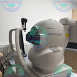
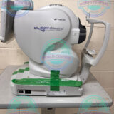
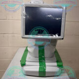
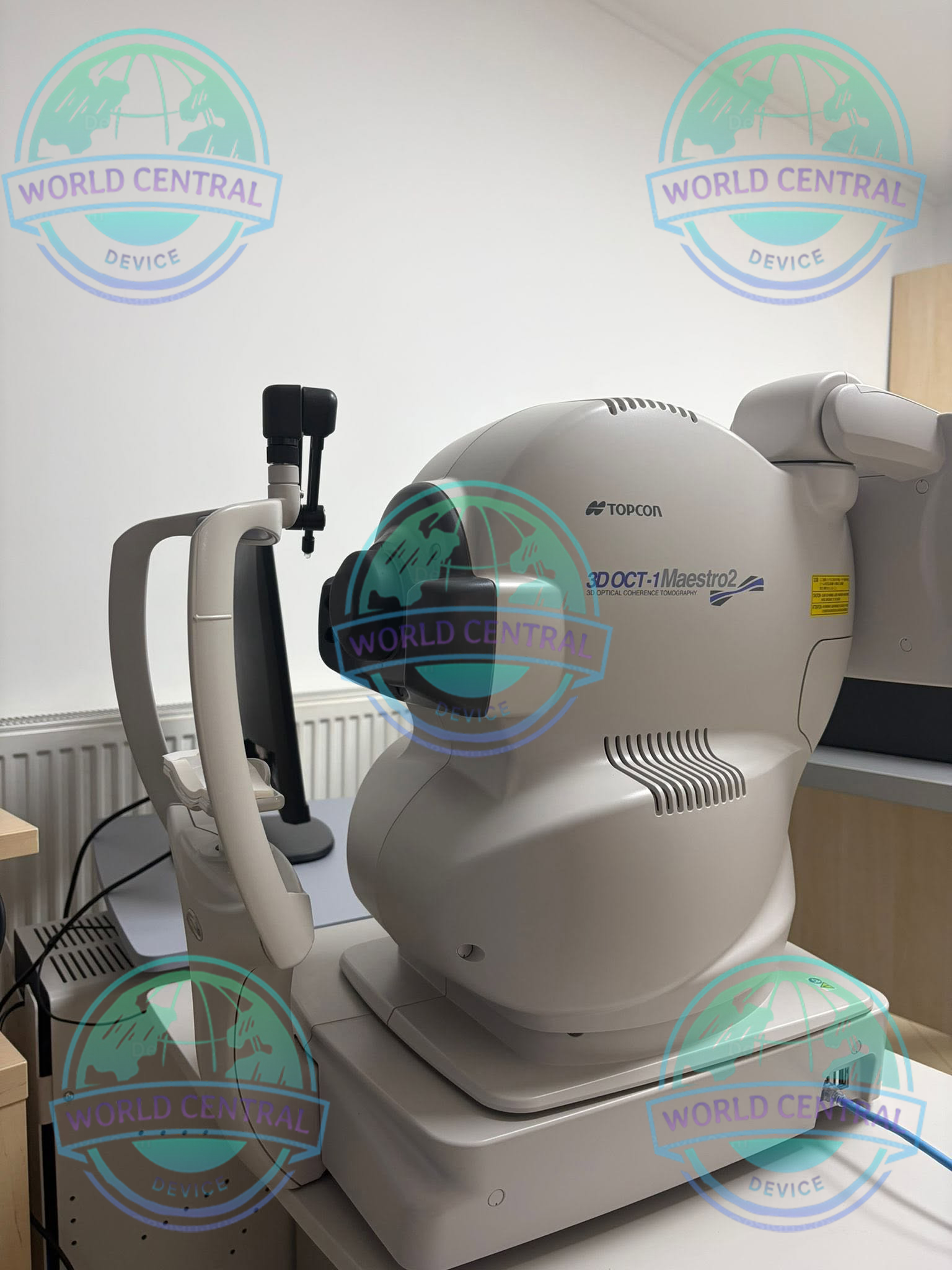
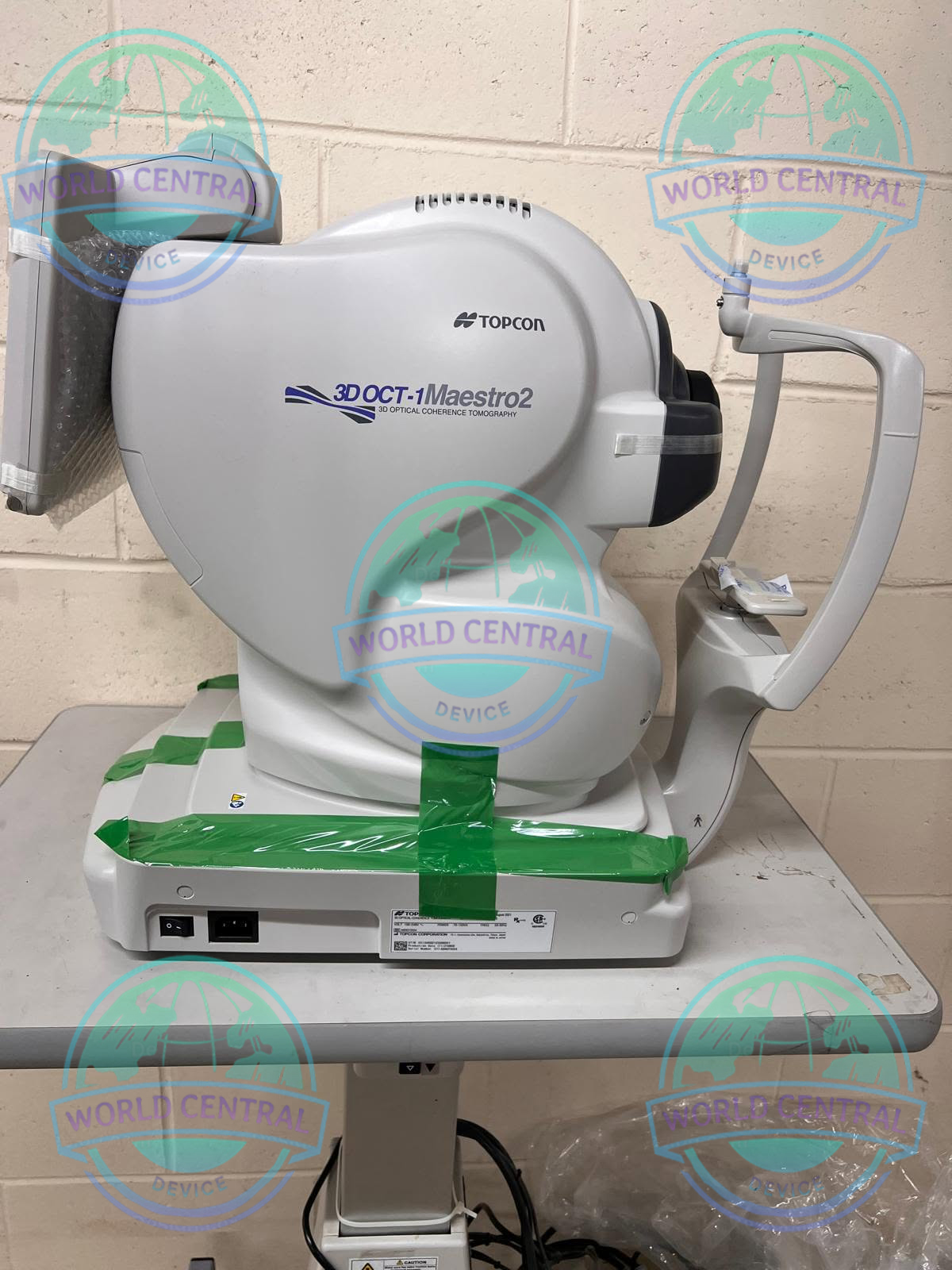
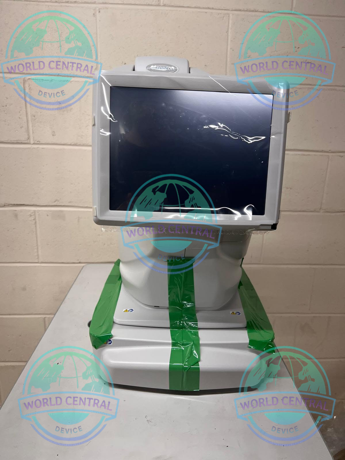
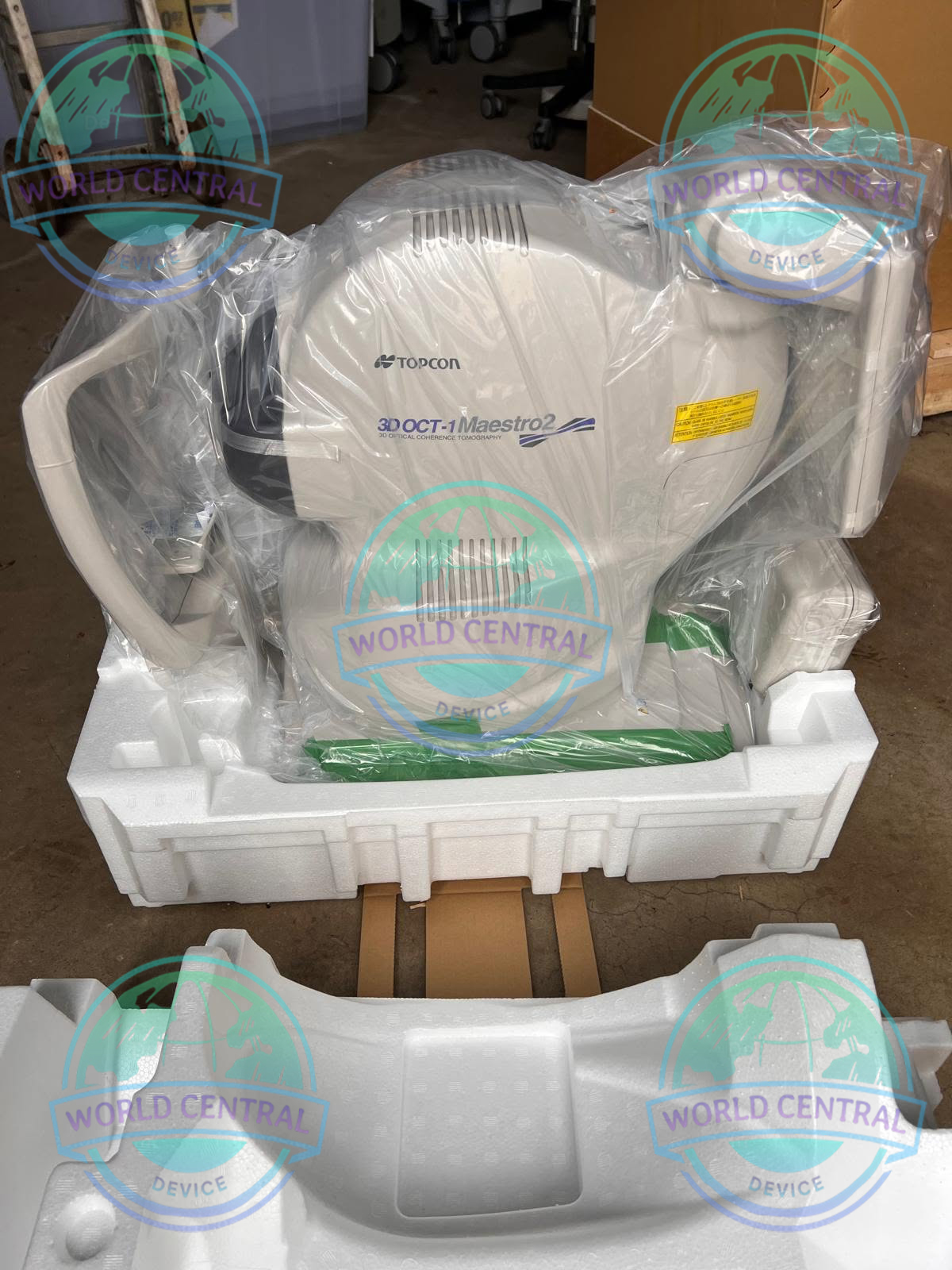
Topcon 3D OCT-1 Maestro 2 OCT System
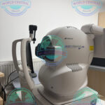
The Topcon 3D OCT-1 Maestro 2 OCT System is an advanced Spectral-Domain OCT featuring fully automated robotic image acquisition, integrated true-color fundus photography, widefield retinal scanning, optional OCT Angiography (OCTA), and comprehensive glaucoma analysis. Designed for ophthalmology clinics
Delivery and return
Delivery and return
Shipping Information
Shipping Information
Composition and care
Composition and care
Description
Topcon 3D OCT-1 Maestro 2 OCT System
Advanced Robotic OCT & True Color Fundus Imaging for Modern Eye Care
The Topcon 3D OCT-1 Maestro 2 OCT System is an advanced Spectral-Domain Optical Coherence Tomography (SD-OCT) platform that combines robotic OCT scanning, true-color non-mydriatic fundus photography, and optional OCT Angiography (OCTA) into a single compact workstation. Designed to improve clinical workflow and diagnostic confidence, Maestro 2 performs automatic alignment, focusing, optimization, and image acquisition with a single touch.
Built for ophthalmologists, retina specialists, glaucoma clinics, optometrists, and eye hospitals, the Maestro 2 enables high-quality retinal imaging while minimizing operator dependency. Its intuitive touchscreen interface and automated capture make it ideal for both experienced clinicians and high-volume screening environments.
The integrated imaging technology provides simultaneous structural OCT data and true-color retinal photography, allowing clinicians to accurately correlate retinal pathology with anatomical landmarks for faster and more reliable diagnosis.
Why Choose Topcon 3D OCT-1 Maestro 2?
Modern ophthalmic practices require fast, reliable imaging without sacrificing diagnostic accuracy. The Maestro 2 delivers a fully automated workflow that simplifies examinations while providing comprehensive retinal analysis.
Key advantages include:
- Fully robotic one-touch image acquisition
- Spectral-Domain OCT technology
- Integrated True Color Fundus Camera
- Optional OCT Angiography (OCTA)
- Widefield 12 × 9 mm OCT scan
- Automatic retinal layer segmentation
- Hood Report for glaucoma evaluation
- RNFL, GCL+, GCL++, and retinal thickness analysis
- Small pupil imaging capability
- Compact footprint for smaller examination rooms
- DICOM and network compatibility
- Optional anterior segment OCT imaging
Key Features
Fully Automated Robotic OCT
Maestro 2 automates nearly every step of image acquisition including:
- Auto Alignment
- Auto Focus
- Auto Optimization
- Auto Capture
Technical Details
Angle of photography: 45° ± 5% or less
Scanning speed: 50,000 A-Scans per second
Working distance: 34.8 ± 0.1mm (for fundus photography) – 62.6 ± 0.1mm (for anterior segment photography)
Pupil diameter: Normal pupil diameter: ø4.0mm or more; Small pupil diameter: ø3.3mm or more
The Maestro2 acquires a 12 mm x 9 mm wide-field 3D OCT, ideal for annual follow-up. It offers features such as the Hood Report for glaucoma and a probability map with 3D Wide 12x9mm acquisition. OCT Angiography is also fully automated, providing information on vascular structures without contrast injection.
True Color Fundus Photography
Unlike pseudo-color imaging systems, Maestro 2 captures genuine 24-bit true-color retinal photographs simultaneously with OCT scans.
Benefits include:
- Accurate retinal documentation
- Precise pathology localization
- PinPoint registration between OCT and fundus image
- Improved follow-up comparison
Widefield OCT Imaging
The large 12 × 9 mm Wide Scan captures:
- Macula
- Optic Disc
- RNFL
within a single acquisition, reducing patient chair time while expanding diagnostic coverage.
Hood Glaucoma Report
Developed around the concepts introduced by Professor Donald Hood, the Hood Report provides simplified visualization of glaucoma progression by correlating structural OCT findings with visual field locations.
Ideal for:
- Glaucoma diagnosis
- Disease progression monitoring
- Clinical decision support
OCT Angiography (Optional)
The optional OCTA module visualizes retinal and choroidal microvasculature without dye injection.
Applications include:
- Diabetic Retinopathy
- Retinal Vein Occlusion
- AMD
- Macular Ischemia
- Vascular abnormalities
Automatic Retinal Layer Segmentation
The software automatically analyzes:
- Retina Thickness
- RNFL Thickness
- GCL+
- GCL++
- Optic Disc Parameters
Anterior Segment OCT
Using the optional anterior segment attachment, clinicians can evaluate:
- Cornea
- Anterior Chamber
- Iris
- Lens
without changing imaging systems.
Clinical Applications
Retina
- Age-related Macular Degeneration (AMD)
- Diabetic Macular Edema
- Diabetic Retinopathy
- Macular Hole
- Epiretinal Membrane
- Central Serous Retinopathy
- Retinal Vein Occlusion
Glaucoma
- RNFL analysis
- Ganglion Cell Complex evaluation
- Optic Nerve Head assessment
- Progression monitoring
- Hood Report
General Ophthalmology
- Routine eye examinations
- Screening programs
- Follow-up imaging
- Documentation
Anterior Segment
- Corneal evaluation
- Anterior chamber imaging
- Post-operative assessment
Technical Specifications
| Category | Specification |
|---|---|
| Product Name | Topcon 3D OCT-1 Maestro 2 |
| Imaging Technology | Spectral-Domain Optical Coherence Tomography (SD-OCT) |
| OCT Type | Spectral Domain |
| Integrated Camera | True Color Non-Mydriatic Fundus Camera |
| Optional Module | OCT Angiography (OCTA) |
| Capture Mode | Fully Automatic / Manual / Semi-Automatic |
| Auto Functions | Auto Alignment, Auto Focus, Auto Optimization, Auto Capture |
| Scan Speed | 50,000 A-Scans/sec |
| Axial Resolution | ≤6 μm |
| Lateral Resolution | ≤20 μm |
| OCT Scan Width | 3–12 mm |
| OCT Scan Height | 3–9 mm |
| Wide Scan | 12 × 9 mm |
| Scan Patterns | 3D Volume Scan, Horizontal, Vertical, Line Scan, Cross Scan, Radial Scan |
| Fundus Photography | True Color |
| Additional Imaging | Digital Red-Free |
| Infrared Imaging | Yes |
| Image Angle | 45° ±5% |
| Digital Zoom | Equivalent 30° |
| Fundus Working Distance | 34.8 ±0.1 mm |
| Anterior Segment Imaging | Optional |
| Corneal Scan Range | 3–6 mm |
| Corneal Scan Patterns | Line Scan, Radial Scan |
| Internal Fixation Target | OLED Dot Matrix |
| External Fixation Target | Yes |
| Minimum Pupil Size (Fundus) | 3.3 mm |
| Minimum Pupil Size (OCT) | 2.5 mm |
| Automatic Layer Segmentation | Yes |
| Retina Thickness Analysis | Yes |
| RNFL Analysis | Yes |
| Ganglion Cell Analysis | GCL+, GCL++ |
| Optic Disc Analysis | Yes |
| Hood Glaucoma Report | Yes |
| Normative Database | Retina, RNFL, GCL+, GCL++ |
| PinPoint Registration | Yes |
| Simultaneous OCT & Fundus | Yes |
| Follow-Up Registration | Yes |
| Touchscreen Monitor | Full Rotating Touchscreen |
| Image Export | DICOM Compatible |
| Network Support | Yes |
| EMR Integration | Supported |
| Software Reports | Retina, Glaucoma, RNFL, Macula |
| Clinical Applications | Retina, Glaucoma, General Ophthalmology, Screening |
| Device Footprint | Compact |


There are no reviews yet.