
No products in the cart.: $0.00

On Sale!


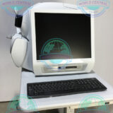
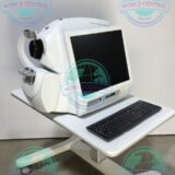
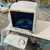
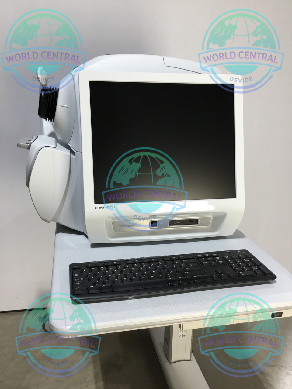
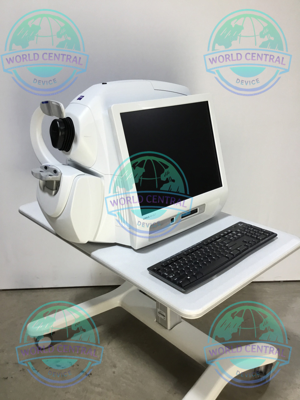
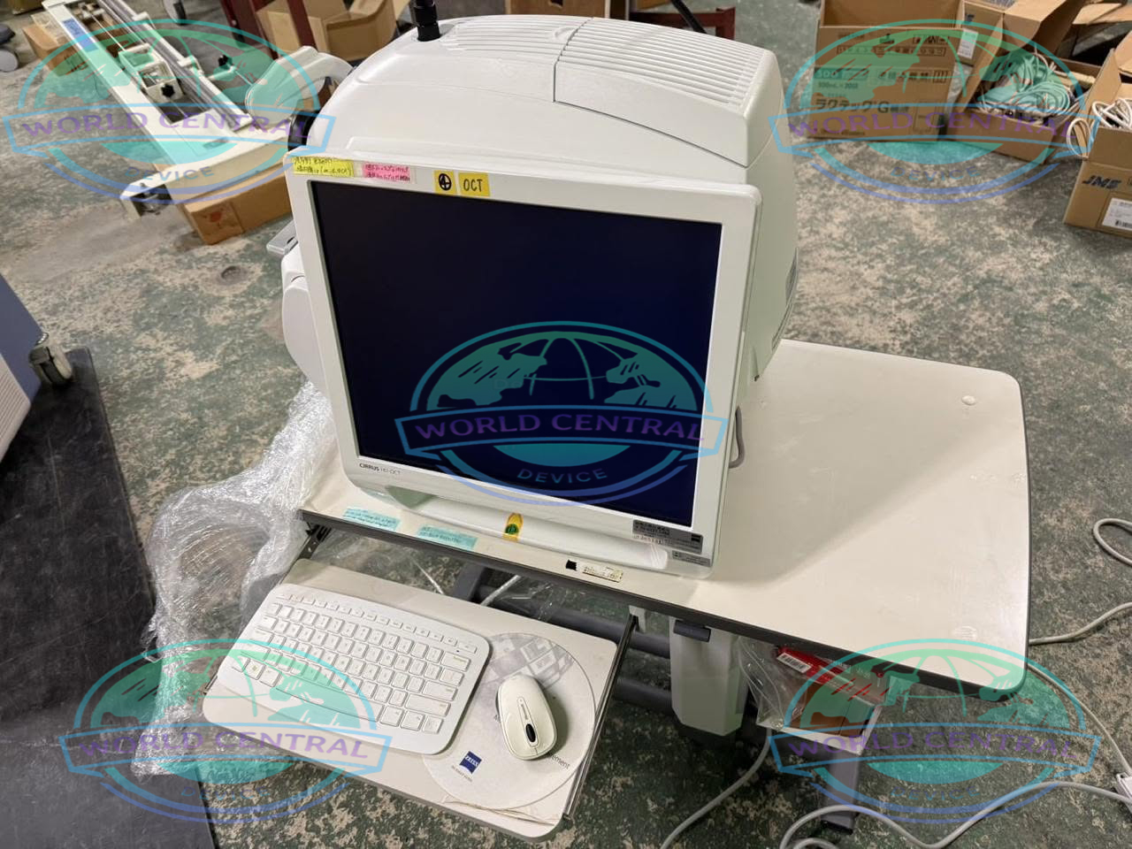
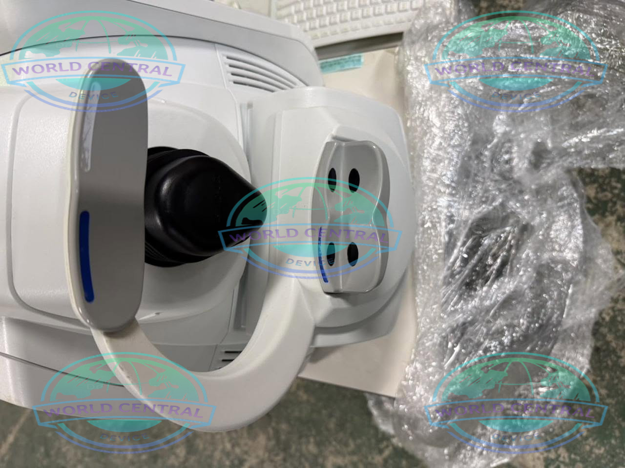
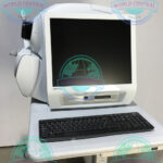
ZEISS CIRRUS 5000 OCT System
The ZEISS CIRRUS 5000 OCT System is a premium Spectral Domain Optical Coherence Tomography (SD-OCT) platform designed for high-resolution imaging of the retina, macula, optic nerve, and anterior segment. Equipped with FastTrac™ retinal tracking, Ganglion Cell Analysis (GCA), RNFL analysis
Delivery and return
Delivery and return
Shipping Information
Shipping Information
Composition and care
Composition and care
Description
ZEISS CIRRUS 5000 OCT System
Advanced Spectral Domain Optical Coherence Tomography for Comprehensive Retinal, Glaucoma, and Anterior Segment Imaging
The ZEISS CIRRUS 5000 OCT System is a premium Spectral Domain Optical Coherence Tomography (SD-OCT) platform designed to deliver high-resolution cross-sectional imaging of the retina, optic nerve, macula, and anterior segment. Built upon ZEISS’ renowned imaging technology, the CIRRUS 5000 combines exceptional image quality with advanced clinical analytics to support early disease detection, treatment planning, and long-term patient monitoring.
Designed for ophthalmologists, retina specialists, glaucoma clinics, hospitals, and eye care centers, the system offers fast image acquisition, intelligent workflow automation, and integrated software that enhances diagnostic confidence. With its FastTrac™ retinal tracking technology, robust normative databases, and advanced progression analysis, the CIRRUS 5000 is an ideal solution for comprehensive ophthalmic imaging.
Why Choose the ZEISS CIRRUS 5000 OCT System?
Selecting the right OCT platform is essential for accurate diagnosis and efficient patient management. The ZEISS CIRRUS 5000 has earned worldwide recognition for its precision, repeatability, and clinical reliability.
Its advanced imaging capabilities allow clinicians to visualize retinal microstructures in remarkable detail while providing quantitative measurements that support evidence-based decision-making.
Key advantages include:
- High-resolution Spectral Domain OCT imaging
- Fast image acquisition with minimal motion artifacts
- Intelligent retinal tracking technology
- Comprehensive glaucoma assessment
- Advanced macular disease evaluation
- Optional anterior segment imaging
- Wide range of scan protocols
- Powerful progression analysis software
- Intuitive touchscreen interface
- DICOM compatibility for hospital integration
- Reliable follow-up registration
- Trusted ZEISS optical engineering
What is Optical Coherence Tomography (OCT)?
Understanding OCT Technology
Optical Coherence Tomography (OCT) is a non-invasive imaging modality that uses low-coherence interferometry to generate high-resolution cross-sectional images of ocular tissues.
Unlike conventional fundus photography, OCT provides depth-resolved visualization of retinal layers, allowing clinicians to detect subtle structural abnormalities that may not be visible during routine examination.
The ZEISS CIRRUS 5000 utilizes Spectral Domain OCT technology to achieve rapid image acquisition with exceptional axial resolution, enabling early detection of retinal and optic nerve pathology.
Benefits of Spectral Domain OCT
Spectral Domain OCT offers several advantages over earlier time-domain systems.
These include:
- Faster scan speeds
- Higher image resolution
- Improved reproducibility
- Reduced motion artifacts
- Better visualization of retinal microstructures
- Enhanced progression analysis
- Improved patient comfort
- Increased diagnostic confidence
Key Features of the ZEISS CIRRUS 5000 OCT System
High-Speed Spectral Domain Imaging
The CIRRUS 5000 captures high-density retinal scans in just seconds, allowing clinicians to obtain detailed structural information while minimizing patient movement.
Rapid scanning enhances workflow efficiency and improves image consistency during routine examinations.
Features
PanoMap Wide-Field Analysis
Integrated Insights
Backward Compatibility
Full Anterior Chamber Imaging
HD Cornea Scan
Pachymetry Map
Wide Angle-to-Angle Scan and HD Angle Scan
Smart HD 1 Line Scan
VRI en Face Preset Display
IS/OS-Ellipsoid en Face Preset Display
FastTrac™ Retinal Tracking Technology
FastTrac technology compensates for involuntary eye movement during image acquisition.
Benefits include:
- Improved scan registration
- More accurate follow-up imaging
- Better progression analysis
- Reduced motion artifacts
- Higher reproducibility
HD-OCT Imaging
High-definition scanning produces exceptional visualization of retinal layers, enabling clinicians to detect subtle pathological changes associated with retinal diseases.
The detailed cross-sectional images support accurate diagnosis and treatment monitoring.
Macular Cube Analysis
Macular Cube scanning provides comprehensive assessment of the macular region by generating volumetric datasets.
Clinical measurements include:
- Macular thickness
- Retinal volume
- ETDRS grid analysis
- Thickness deviation maps
- Thickness progression
Optic Disc Cube Analysis
The Optic Disc Cube protocol evaluates the optic nerve head and retinal nerve fiber layer (RNFL).
It provides:
- RNFL thickness maps
- Quadrant analysis
- Clock-hour analysis
- Optic nerve head parameters
- Disc area measurements
- Cup-to-disc ratio
- Rim area evaluation
Ganglion Cell Analysis (GCA)
Ganglion Cell Analysis enables early glaucoma detection by measuring the ganglion cell-inner plexiform layer (GCIPL).
Clinical outputs include:
- GCIPL thickness
- Sector analysis
- Deviation maps
- Thickness asymmetry
- Progression monitoring
Advanced Progression Analysis
Longitudinal comparison tools automatically align previous examinations to monitor disease progression.
Clinicians can identify structural changes over time with improved confidence.
Anterior Segment Imaging
With the appropriate lens configuration, the CIRRUS 5000 can image anterior ocular structures, including:
- Cornea
- Iris
- Anterior chamber
- Angle structures
- Lens
Clinical Applications
Retina Imaging
The ZEISS CIRRUS 5000 supports diagnosis and monitoring of numerous retinal diseases.
Common applications include:
- Age-related macular degeneration (AMD)
- Diabetic macular edema (DME)
- Diabetic retinopathy
- Epiretinal membrane
- Macular hole
- Vitreomacular traction
- Retinal edema
- Retinal detachment assessment
- Central serous chorioretinopathy
- Retinal vascular disorders
Glaucoma Assessment
The integrated glaucoma analysis tools assist clinicians in detecting early structural damage before significant visual field loss occurs.
Assessments include:
- RNFL analysis
- Optic nerve head evaluation
- Ganglion cell analysis
- Progression monitoring
- Normative database comparison
Neuro-Ophthalmology
The CIRRUS 5000 assists in evaluating optic nerve disorders such as:
- Optic neuritis
- Ischemic optic neuropathy
- Papilledema
- Optic atrophy
- Multiple sclerosis-related optic nerve damage
Anterior Segment Evaluation
Clinicians can visualize anterior ocular structures for:
- Corneal assessment
- Angle evaluation
- Refractive surgery planning
- Postoperative follow-up
- Contact lens assessment
Imaging Technology
Spectral Domain OCT Engine
The CIRRUS 5000 employs Spectral Domain OCT technology to produce high-speed volumetric retinal imaging with excellent axial resolution.
Its optimized optical design ensures consistent image quality while reducing acquisition time.
Intelligent Scan Registration
Automatic scan registration improves follow-up examinations by aligning current scans with previous datasets.
This enables highly reliable progression analysis for glaucoma and retinal diseases.
Advanced Image Processing
Sophisticated image processing algorithms enhance:
- Layer segmentation
- Noise reduction
- Retinal boundary detection
- Quantitative measurements
- Visualization quality
General Specifications
| Parameter | Specification |
|---|---|
| Product Name | ZEISS CIRRUS 5000 OCT System |
| Manufacturer | ZEISS |
| Device Type | Spectral Domain Optical Coherence Tomography (SD-OCT) System |
| Imaging Technology | Spectral Domain Optical Coherence Tomography |
| Intended Use | Diagnostic ophthalmic imaging |
| Clinical Applications | Retina, Macula, Glaucoma, Optic Nerve Head, RNFL, Ganglion Cell Analysis, Anterior Segment (optional) |
| Installation Type | Tabletop / Desktop |
| Operation | Computer-controlled |
| User Interface | Windows-based graphical interface |
| Patient Position | Sitting |
| Alignment Method | Motorized alignment with joystick |
| Fixation | Internal fixation target |
| External Fixation | Optional |
| Target Patient Population | Pediatric and adult patients (clinician dependent) |
| Image Acquisition | Non-contact |
| Scan Registration | Automatic |
| Follow-up Registration | Automatic |
Optical Specifications
| Parameter | Specification |
|---|---|
| OCT Technology | Spectral Domain OCT |
| Light Source Type | Superluminescent Diode (SLD) |
| Central Wavelength | Approximately 840 nm |
| Spectral Bandwidth | Approximately 50 nm |
| Axial Resolution (Tissue) | Approximately 5 µm |
| Axial Resolution (Air) | Approximately 3–4 µm |
| Transverse Resolution | Approximately 15–20 µm |
| Optical Focus | Automatic |
| Optical Alignment | Motorized |
| Optical Magnification | Software compensated |
| Imaging Beam Power | Within international ophthalmic safety standards |
| Eye Safety Classification | Class 1 ophthalmic laser product |
| Maximum Exposure | ANSI/IEC compliant |
| Scan Depth | Approximately 2.0 mm |
| Dynamic Range | High dynamic range imaging |
| Optical Distortion Correction | Automatic |
| Optical Calibration | Factory calibrated |
Imaging Performance
| Parameter | Specification |
|---|---|
| A-Scan Speed | 27,000 A-scans/second |
| Scan Acquisition Time | Approximately 1–2 seconds |
| Maximum B-Scan Density | High-density acquisition |
| B-Scan Display | Real-time |
| Scan Averaging | Supported |
| Motion Artifact Reduction | FastTrac™ |
| Eye Motion Compensation | Yes |
| Signal Strength Indicator | Yes |
| Image Enhancement | Automatic |
| Image Optimization | Automatic |
| Noise Reduction | Integrated |
| Layer Segmentation | Automatic |
| Manual Segmentation Correction | Yes |
| Thickness Mapping | Yes |
| Reflectivity Mapping | Yes |
| En Face Visualization | Supported (software version dependent) |
| Image Export Format | JPEG |
| Additional Export | TIFF |
| Report Export | |
| Raw Data Archive | Supported |

There are no reviews yet.