
No products in the cart.: $0.00

On Sale!


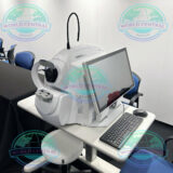
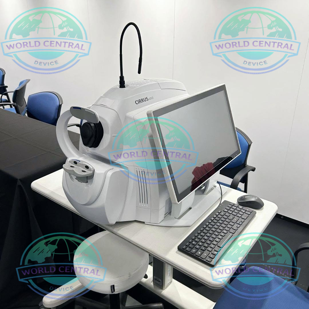
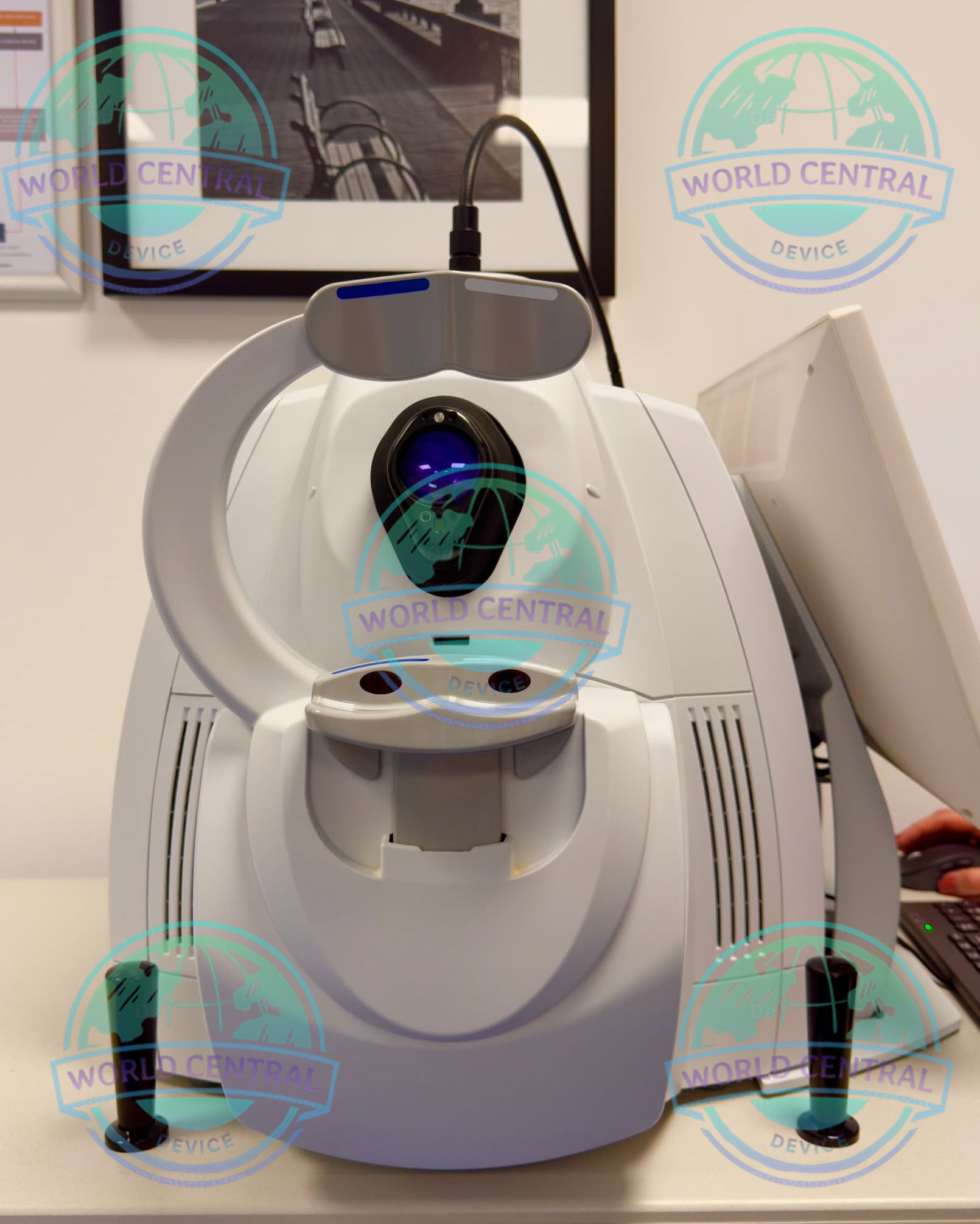
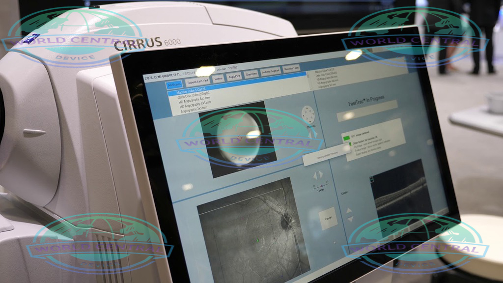
ZEISS CIRRUS 6000 OCT System
The ZEISS CIRRUS 6000 OCT System is a premium Spectral Domain Optical Coherence Tomography (SD-OCT) platform engineered for high-resolution retinal imaging and comprehensive ophthalmic diagnostics. Equipped with FastTrac™ eye tracking, AngioPlex™ OCT Angiography, Ganglion Cell Analysis (GCA), Retinal Nerve Fiber Layer (RNFL) analysis
Delivery and return
Delivery and return
Shipping Information
Shipping Information
Composition and care
Composition and care
Description
ZEISS CIRRUS 6000 OCT System
Advanced Spectral Domain OCT for High-Speed Retinal Imaging and Comprehensive Ophthalmic Diagnostics
The ZEISS CIRRUS 6000 OCT System is a next-generation Spectral Domain Optical Coherence Tomography (SD-OCT) platform designed to deliver exceptional image quality, high-speed scanning, and advanced retinal analysis for comprehensive ophthalmic diagnostics. Built on ZEISS optical engineering, the CIRRUS 6000 combines rapid image acquisition, intelligent eye tracking, and sophisticated analytical software to support the diagnosis and long-term management of retinal diseases, glaucoma, and optic nerve disorders.
Engineered for ophthalmologists, retina specialists, glaucoma clinics, hospitals, and research institutions, the CIRRUS 6000 provides detailed structural imaging, OCT angiography, and automated progression analysis in a single, user-friendly platform. Its advanced workflow features help improve clinical efficiency while delivering reliable and reproducible diagnostic results.
Why Choose the ZEISS CIRRUS 6000 OCT System?
The ZEISS CIRRUS 6000 is designed to meet the growing demand for faster examinations, higher image quality, and more comprehensive ocular analysis. By integrating advanced imaging technologies with intelligent software, it enables clinicians to detect subtle structural changes at an early stage and monitor disease progression with confidence.
Key Advantages
- High-speed Spectral Domain OCT imaging
- Advanced AngioPlex™ OCT Angiography
- FastTrac™ retinal eye tracking technology
- High-definition retinal and optic nerve imaging
- Automated retinal layer segmentation
- Ganglion Cell Analysis (GCA)
- Retinal Nerve Fiber Layer (RNFL) analysis
- Optic Nerve Head (ONH) assessment
- Guided Progression Analysis (GPA)
- Comprehensive glaucoma evaluation
- DICOM and EMR integration
- Intuitive software with customizable reports
Features
High-Speed Imaging: 100,000 A-scans per second, with OCT cube scans completed in 0.4 seconds.
Wide Field of View: Large scan areas up to 12×12 mm for a comprehensive retinal view.
Enhanced Microvascular Detail: HD AngioPlex scans with 8×8 and 6×6 mm field sizes.
Proven Analytics: Clinically-validated tools for retina, glaucoma, and anterior segment analysis.
Seamless Data Transfer: Compatibility with previous CIRRUS models for continuity of care.
Motion Artifact Reduction: FastTrac eye tracking technology ensures crisp, detailed images.
Performance OCT — faster, wider, with a new level of detail
ZEISS CIRRUS 6000 empowers clinicians with a larger field of view in a single scan, and captures high-definition OCT/OCTA scans that reveal finer details of the retinal microvasculature − all of which provides more insight into the patient’s condition in less time.
Proven analytics: CIRRUS-powered treatment decisions
As the pioneering OCT technology, the CIRRUS platform offers clinicians extensive, clinically-validated applications for retina, glaucoma and anterior segment. The result: precise analysis, faster throughput and smarter decision-making across a wide spectrum of clinical conditions and patient types.
Retina
Macular Change Analysis The CIRRUS data cube automatically stores and delivers each patient’s historical data to provide a variety of change assessments, including macular thickness change maps that help you understand your patient‘s response to treatment. Because every CIRRUS cube is tracked and registered to OCT scans from prior visits using CIRRUS’ FastTrac™ Retinal Tracking Technology, you can confidently measure point-to-point changes in macular thickness.
Spectral Domain OCT Technology
High-Speed Image Acquisition
The CIRRUS 6000 utilizes Spectral Domain OCT (SD-OCT) technology to acquire high-resolution cross-sectional images of ocular tissues within seconds. The fast scanning speed minimizes motion artifacts and improves patient comfort during examinations.
Benefits of SD-OCT
- Rapid image acquisition
- Excellent axial resolution
- Detailed retinal layer visualization
- Reliable follow-up imaging
- Improved diagnostic confidence
- Enhanced workflow efficiency
AngioPlex™ OCT Angiography
Non-Invasive Retinal Microvasculature Imaging
The integrated AngioPlex™ OCT Angiography module provides dye-free visualization of retinal and choroidal blood vessels. This technology allows clinicians to evaluate retinal microcirculation without the need for fluorescein dye injection.
Clinical Applications
- Diabetic Retinopathy
- Age-related Macular Degeneration (AMD)
- Retinal Vein Occlusion (RVO)
- Choroidal Neovascularization (CNV)
- Macular Ischemia
- Vascular Abnormalities
Retinal Imaging
High-Definition Retinal Scans
The CIRRUS 6000 captures high-definition B-scan images with exceptional clarity, enabling clinicians to evaluate retinal anatomy layer by layer.
Common Retinal Assessments
- Macular Thickness
- Retinal Volume
- Retinal Layer Segmentation
- Epiretinal Membrane (ERM)
- Macular Hole
- Vitreomacular Traction (VMT)
- Cystoid Macular Edema (CME)
Ganglion Cell Analysis (GCA)
The integrated Ganglion Cell Analysis software measures the ganglion cell-inner plexiform layer (GCIPL), supporting the early detection and monitoring of glaucoma.
Parameters Evaluated
- Average GCIPL Thickness
- Minimum GCIPL Thickness
- Sectoral Thickness
- Deviation Maps
- Thickness Maps
- Asymmetry Analysis
RNFL Analysis
Comprehensive Glaucoma Evaluation
The Retinal Nerve Fiber Layer (RNFL) analysis module provides detailed measurements of nerve fiber thickness surrounding the optic disc.
Features
- Average RNFL Thickness
- Quadrant Analysis
- Clock Hour Analysis
- Deviation Mapping
- Normative Database Comparison
- Progression Monitoring
Optic Nerve Head (ONH) Analysis
The ONH analysis module evaluates optic disc morphology using automated measurements, assisting clinicians in glaucoma diagnosis and disease progression monitoring.
Measured parameters include:
- Disc Area
- Rim Area
- Cup Volume
- Cup-to-Disc Ratio
- Vertical Cup-to-Disc Ratio
- Horizontal Cup-to-Disc Ratio
FastTrac™ Eye Tracking
FastTrac™ retinal tracking technology continuously compensates for eye movement during image acquisition, ensuring consistent image registration and improved repeatability between follow-up examinations.
Benefits
- Reduced motion artifacts
- Improved scan alignment
- Reliable progression analysis
- Enhanced measurement consistency
Clinical Applications
The ZEISS CIRRUS 6000 OCT System supports a broad range of ophthalmic diagnostic applications, including:
Retina
- Age-related Macular Degeneration (AMD)
- Diabetic Macular Edema (DME)
- Diabetic Retinopathy (DR)
- Epiretinal Membrane (ERM)
- Macular Hole
- Central Serous Chorioretinopathy (CSC)
- Retinal Vein Occlusion (RVO)
Glaucoma
- Early Glaucoma Detection
- RNFL Analysis
- Ganglion Cell Analysis
- Optic Nerve Assessment
- Guided Progression Analysis
Neuro-Ophthalmology
- Optic Neuropathy Evaluation
- Papilledema Monitoring
- Optic Disc Edema Assessment
Cataract & Refractive Surgery
- Preoperative Retinal Screening
- Macular Assessment Before Cataract Surgery
- Premium IOL Evaluation
Benefits for Clinical Practice
Enhanced Diagnostic Confidence
High-resolution imaging and automated analysis help clinicians detect subtle structural abnormalities earlier, supporting timely diagnosis and treatment.
Improved Workflow Efficiency
Fast acquisition, intelligent eye tracking, and automated reporting reduce examination time and improve patient throughput.
Comprehensive Data Analysis
Integrated software combines retinal imaging, glaucoma metrics, angiography, and progression analysis into a unified diagnostic platform.
Reliable Long-Term Monitoring
Advanced follow-up registration ensures consistent comparisons between visits, enabling accurate assessment of disease progression.
Key Features at a Glance
- Spectral Domain OCT Technology
- AngioPlex™ OCT Angiography
- FastTrac™ Eye Tracking
- High-Speed Image Acquisition
- High-Definition B-Scan Imaging
- Automated Retinal Layer Segmentation
- Ganglion Cell Analysis (GCA)
- RNFL Thickness Analysis
- Optic Nerve Head Assessment
- Guided Progression Analysis (GPA)
- Retina Thickness Mapping
- ETDRS Grid Analysis
- DICOM & EMR Connectivity
- User-Friendly Software Interface
Technical specifications
Key Parameters
- Methodology: Spectral domain OCT
- Optical source: Superluminescent diode (SLD), 840 nm
- A-scan depth: 2.0 – 2.9 mm (in tissue)
- Scan speed: 100,000 A-scans per second
- Min. pupil diameter: 2.0 mm
- Resolution:
• Axial resolution
• Transverse resolution
5 μm (in tissue), 1.95 μm (digital)
15 μm (in tissue) - Refractive error adjustment: -20D to +20D (dopters)
- Resolution:
• Axial resolution
• Transverse resolution
5 μm (in tissue), 1.95 μm (digital)
15 μm (in tissue) - Posterior Segment scans:
• OCT
• OCTA
Cube scan (Macula and Optic Disc)
HD Raster (1, 5, 21-line, cross and radial); Raster scan length 3-12 mm;
image averaging up to 100x
3×3, 6×6, 8×8, 12×12 mm (Macula); 4.5×4.5 mm (Optic Nerve Head);
14×10 mm (Montage), 14×14 mm (Montage)


There are no reviews yet.