
No products in the cart.: $0.00

On Sale!


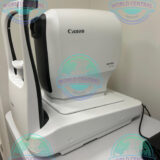
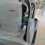
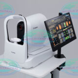
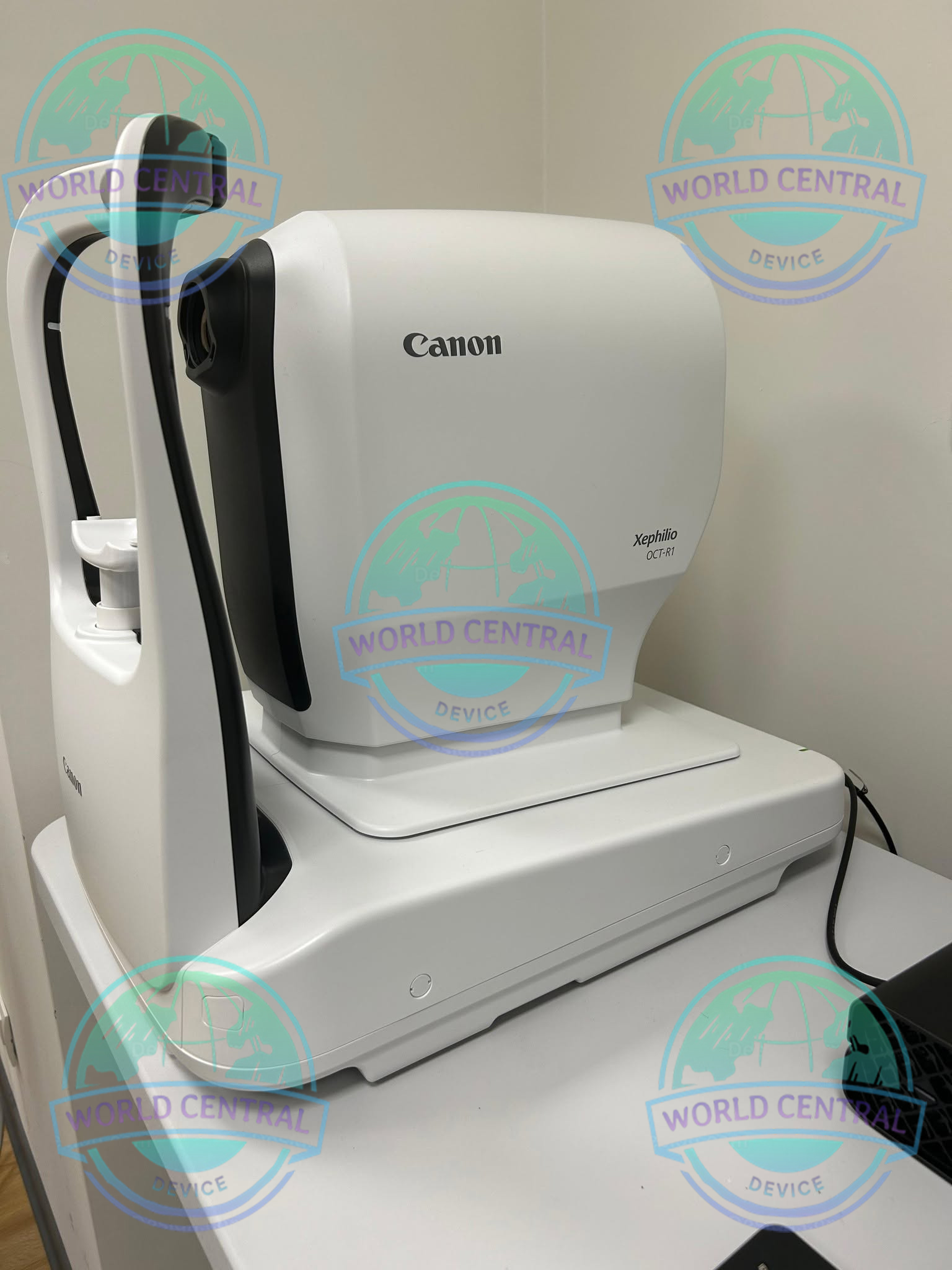
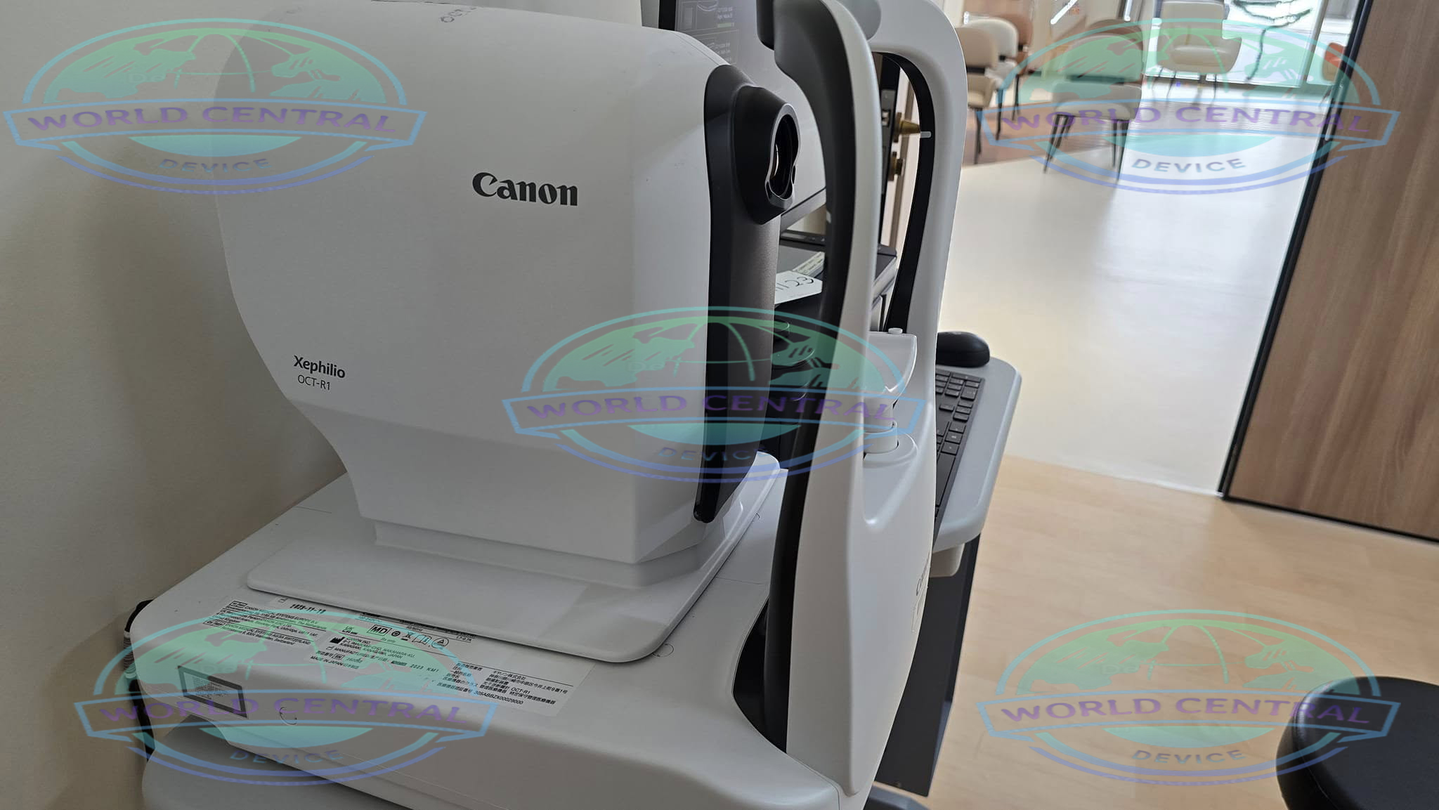
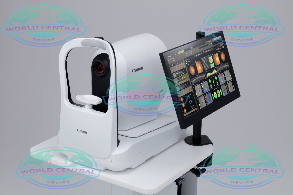
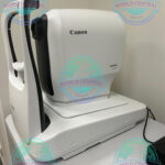
Canon Xephilio OCT-R1
The Canon Xephilio OCT-R1 is an advanced optical coherence tomography (OCT) imaging system designed for comprehensive retinal and optic nerve evaluation. Engineered for ophthalmology clinics and eye care centers, it delivers high-resolution cross-sectional retinal imaging,
Delivery and return
Delivery and return
Shipping Information
Shipping Information
Composition and care
Composition and care
Description
Canon Xephilio OCT-R1
Advanced Optical Coherence Tomography for Comprehensive Retinal Diagnosis
The Canon Xephilio OCT-R1 is an advanced optical coherence tomography (OCT) imaging system developed to support accurate diagnosis, monitoring, and management of retinal and optic nerve diseases. Designed for ophthalmology clinics, hospitals, and eye care centers, the Xephilio OCT-R1 combines high-resolution OCT imaging with intuitive software, efficient workflows, and reliable clinical performance.
By delivering detailed cross-sectional images of retinal structures, the system enables eye care professionals to detect subtle anatomical changes at an early stage. This capability is essential for diagnosing and monitoring conditions such as glaucoma, age-related macular degeneration (AMD), diabetic retinopathy, macular edema, vitreomacular disorders, and other retinal pathologies.
Built with Canon’s expertise in medical imaging, the Xephilio OCT-R1 offers a balance of image quality, user-friendly operation, and streamlined data management. It integrates smoothly into modern ophthalmic practices, helping clinicians improve diagnostic confidence while enhancing patient care.
Why Choose Canon Xephilio OCT-R1?
Designed for Accurate Retinal Imaging
Early diagnosis is one of the most important factors in preserving vision. The Canon Xephilio OCT-R1 provides detailed retinal visualization that assists clinicians in identifying structural abnormalities before significant vision loss occurs.
Key Benefits
- High-resolution retinal imaging
- Detailed cross-sectional OCT scans
- Reliable optic nerve head assessment
- Comprehensive macular analysis
- Efficient glaucoma evaluation
- Intuitive clinical workflow
- Fast patient examination
- High-quality image acquisition
- Advanced analysis software
- Comfortable patient positioning
- User-friendly touchscreen interface
- Digital image storage and management
- Seamless integration into ophthalmic practices
Key Features
High-Resolution OCT Imaging
The Canon Xephilio OCT-R1 produces high-definition cross-sectional images of the retina, allowing clinicians to evaluate individual retinal layers and detect subtle pathological changes.
Benefits include:
- Excellent anatomical detail
- Clear visualization of retinal layers
- Enhanced diagnostic confidence
- Accurate disease monitoring
- Reliable follow-up examinations
Comprehensive Retinal Assessment
The system supports detailed evaluation of multiple retinal conditions through advanced imaging and quantitative analysis.
Common assessment areas include:
- Macular thickness
- Retinal nerve fiber layer (RNFL)
- Optic nerve head
- Ganglion cell complex
- Retinal morphology
- Vitreoretinal interface
Efficient Clinical Workflow
The Xephilio OCT-R1 is designed to improve workflow efficiency by simplifying patient positioning, scan acquisition, and image review.
Workflow Advantages
- Faster examinations
- Reduced operator training
- Consistent imaging quality
- Simplified report generation
- Improved patient throughput
Advanced Analysis Software
Integrated software assists clinicians by providing automated measurements and comparison tools.
Analysis Functions
- Retinal thickness mapping
- RNFL analysis
- Optic disc measurements
- Progression analysis
- Longitudinal patient comparison
- Image review and reporting
Ergonomic Design
The compact and ergonomic system is suitable for both private ophthalmology clinics and large eye hospitals.
Advantages
- Comfortable patient positioning
- Compact footprint
- Easy instrument access
- Intuitive controls
- Efficient operation
Clinical Applications
Retina Evaluation
Assess retinal diseases including diabetic retinopathy, macular degeneration, retinal edema, and vitreomacular interface disorders.
Glaucoma Management
Evaluate retinal nerve fiber layer thickness and optic nerve head morphology for glaucoma diagnosis and progression monitoring.
Macular Disease Assessment
Analyze macular structure to support the diagnosis and treatment of macular disorders.
Diabetic Eye Care
Monitor retinal changes associated with diabetic retinopathy and diabetic macular edema.
Routine Eye Examinations
Support comprehensive ophthalmic examinations through detailed structural imaging.
Long-Term Patient Monitoring
Compare sequential OCT scans to evaluate disease progression and treatment response over time.



There are no reviews yet.