
No products in the cart.: $0.00

On Sale!


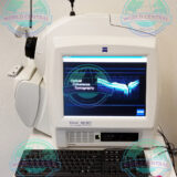
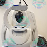
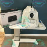
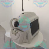
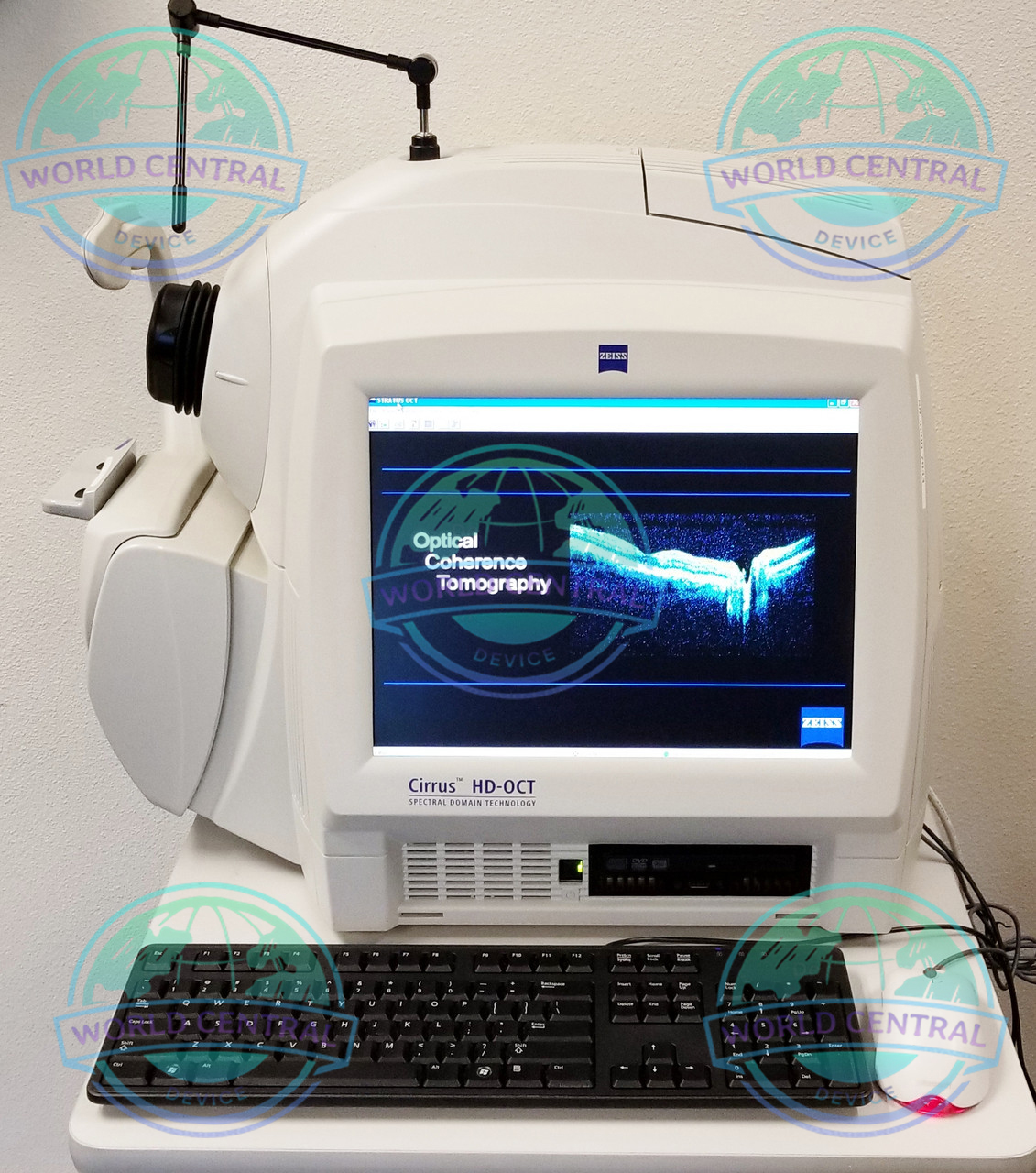
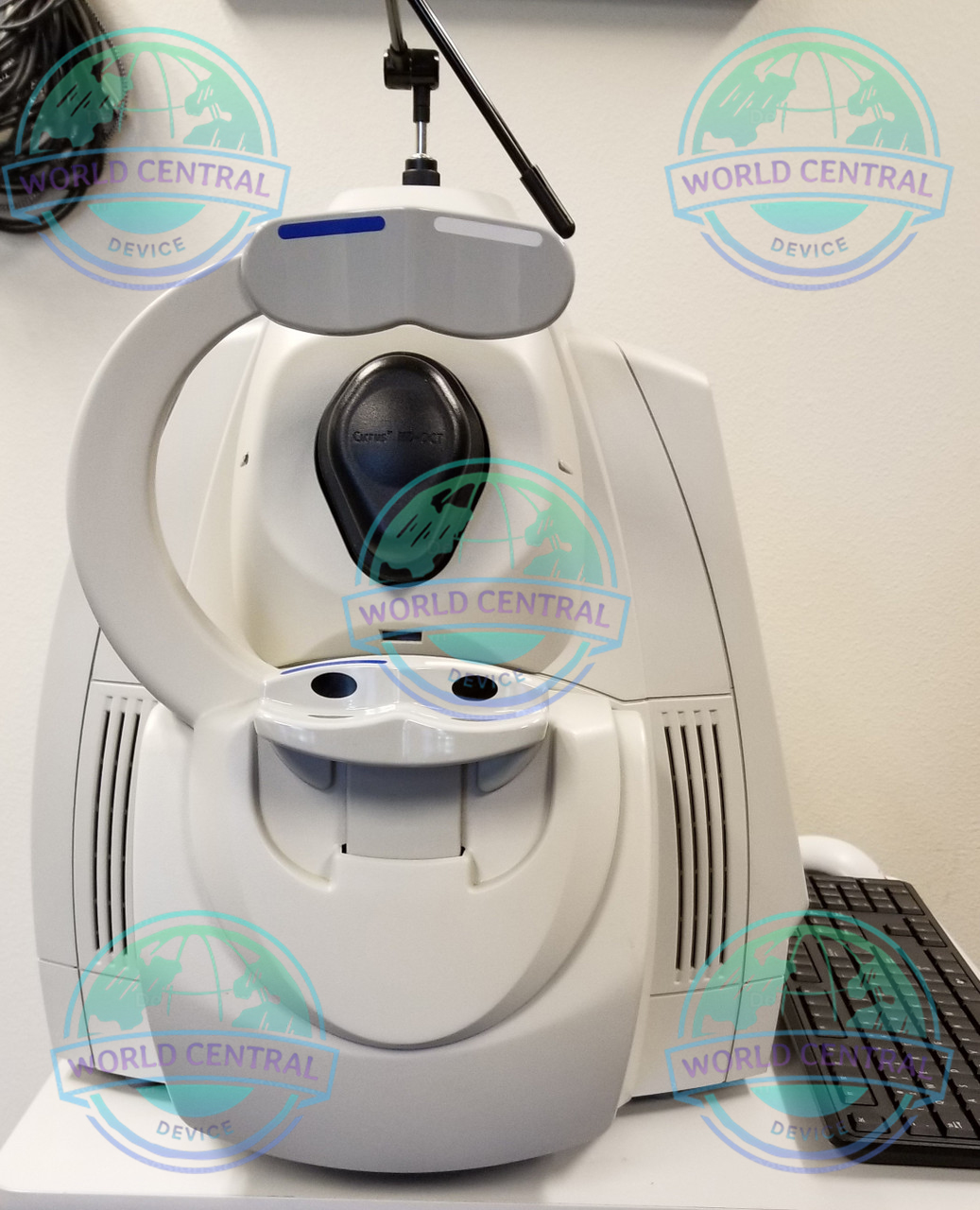
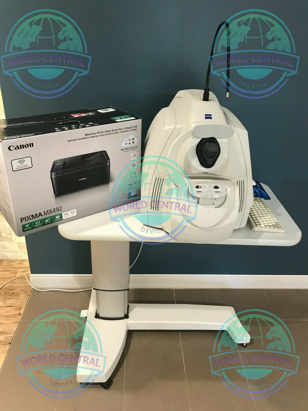
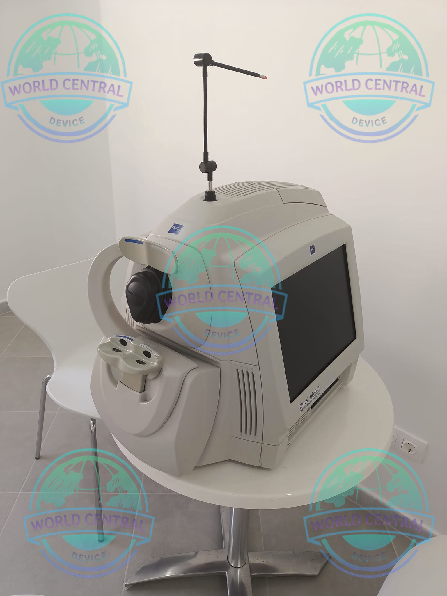
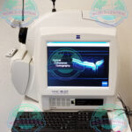
ZEISS CIRRUS 4000 OCT System
The ZEISS CIRRUS 4000 OCT System is a high-performance Spectral Domain Optical Coherence Tomography (SD-OCT) platform designed for comprehensive retinal, optic nerve, and glaucoma imaging. Featuring high-resolution scanning, advanced Ganglion Cell Analysis
Delivery and return
Delivery and return
Shipping Information
Shipping Information
Composition and care
Composition and care
Description
ZEISS CIRRUS 4000 OCT System – High-Resolution Retinal & Optic Nerve Imaging Solution
Advanced Optical Coherence Tomography for Modern Ophthalmology
The ZEISS CIRRUS 4000 OCT System is a powerful optical coherence tomography (OCT) platform designed to provide high-resolution cross-sectional imaging of the retina, optic nerve head, and anterior segment. Developed by ZEISS, this system delivers exceptional image quality, reliable diagnostic data, and streamlined clinical workflow for ophthalmologists, retina specialists, glaucoma specialists, and comprehensive eye care practices.
Built upon proven spectral-domain OCT technology, the CIRRUS 4000 enables clinicians to detect, diagnose, monitor, and manage various ocular diseases with remarkable precision. Whether evaluating glaucoma progression, retinal pathology, or macular disorders, the system produces reproducible measurements that support confident clinical decision-making.
Why Choose the ZEISS CIRRUS 4000 OCT System?
Eye care professionals require imaging systems capable of delivering detailed anatomical information quickly and consistently. The CIRRUS 4000 combines high-speed scanning, intelligent software, and advanced analysis tools into one efficient platform.
Key benefits include:
- Spectral Domain OCT technology
- High-resolution retinal imaging
- Fast acquisition speed
- Excellent repeatability
- Advanced glaucoma analysis
- Macular thickness evaluation
- Optic nerve head assessment
- Ganglion Cell Analysis (GCA)
- Easy patient alignment
- User-friendly software interface
- Reliable longitudinal follow-up
- Comprehensive clinical reporting
Features
High-resolution image acquisition for precise evaluation of the retina and anterior segment of the eye
Capability to visualize and analyze layers of ocular tissues with high precision
Support for multiple acquisition modalities for comprehensive evaluation
User-friendly software platform for intuitive use and efficient image analysis
Compatibility with hospital information systems for seamless integration into existing workflows
Advanced measurement and tracking features for monitoring changes over time
Automated eye alignment system for consistent and reproducible results
Ability to generate detailed and customized reports to facilitate communication with patients and other healthcare professionals
Designed for Comprehensive Eye Disease Management
The CIRRUS 4000 supports diagnosis and monitoring of numerous ophthalmic conditions including:
Glaucoma
Accurate RNFL thickness measurements and optic nerve analysis assist clinicians in detecting glaucoma earlier while monitoring disease progression over time.
Glaucoma Features
- RNFL Thickness Analysis
- Optic Disc Cube Scan
- Ganglion Cell Analysis
- Progression Analysis
- Deviation Maps
- Normative Database Comparison
Retina Disease Assessment
High-resolution macular imaging provides excellent visualization of retinal layers for diagnosing numerous retinal disorders.
Common applications include:
- Age-related Macular Degeneration (AMD)
- Diabetic Macular Edema
- Diabetic Retinopathy
- Epiretinal Membrane
- Macular Hole
- Central Serous Retinopathy
- Retinal Vein Occlusion
- Vitreomacular Traction
Neuro-Ophthalmology
The CIRRUS platform assists neurologists and ophthalmologists in evaluating optic nerve disorders and neurological diseases affecting vision.
Typical examinations include:
- Optic neuritis
- Papilledema
- Multiple sclerosis monitoring
- Optic nerve edema
- Optic nerve atrophy
High-Speed Spectral Domain OCT Technology
The ZEISS CIRRUS 4000 employs Spectral Domain Optical Coherence Tomography (SD-OCT), producing detailed retinal images within seconds.
Advantages include:
- Rapid image acquisition
- Reduced motion artifacts
- Enhanced patient comfort
- Improved scan repeatability
- High-definition retinal visualization
- Excellent layer segmentation
Comprehensive Imaging Capabilities
The CIRRUS 4000 offers multiple scan protocols to accommodate a wide range of clinical examinations.
Posterior Segment Imaging
- Retina
- Macula
- Optic Disc
- RNFL
- Ganglion Cell Layer
- Choroid visualization (limited depending on protocol)
Anterior Segment Imaging (Optional)
With compatible anterior segment lenses, clinicians can visualize:
- Cornea
- Anterior chamber
- Angle structures
- Iris
- Lens
Intelligent Clinical Analysis Software
The integrated software simplifies image review while providing quantitative analysis for disease monitoring.
Software capabilities include:
Thickness Maps
Displays retinal thickness across the macula for rapid pathology detection.
Deviation Maps
Highlights abnormal tissue compared with normative databases.
Progression Analysis
Tracks structural changes across multiple patient visits.
Normative Database
Automatically compares patient measurements with healthy population references.
Comprehensive Reports
Generate professional reports suitable for:
- Clinical documentation
- Electronic Medical Records (EMR)
- Patient consultation
- Referral physicians
Workflow Optimization
The CIRRUS 4000 has been engineered to improve productivity in busy ophthalmology clinics.
Workflow advantages include:
- Fast patient positioning
- Automatic scan acquisition
- Minimal operator training
- Repeatable imaging
- Efficient data management
- Easy follow-up comparisons
Advantages of the ZEISS CIRRUS 4000 OCT System
Compared with conventional retinal imaging systems, the CIRRUS 4000 offers:
- Superior retinal layer visualization
- Reliable quantitative measurements
- Excellent image reproducibility
- Proven ZEISS optical performance
- Comprehensive glaucoma assessment
- Efficient patient throughput
- Intuitive user interface
- Long-term patient monitoring
- Robust clinical database
- Trusted worldwide by eye care professionals
Specifications
| Specification | Details |
|---|---|
| Product Name | ZEISS CIRRUS 4000 OCT System |
| Manufacturer | Carl Zeiss Meditec |
| Technology | Spectral Domain Optical Coherence Tomography (SD-OCT) |
| Imaging Type | Cross-sectional retinal imaging |
| Primary Application | Retina, Optic Nerve, Glaucoma |
| Scan Speed | Up to approximately 27,000 A-scans/second |
| Axial Resolution | Approximately 5 µm in tissue |
| Transverse Resolution | Approximately 15–20 µm |
| Light Source | Superluminescent Diode (SLD) |
| Wavelength | Approximately 840 nm |
| Scan Patterns | Cube Scan, Line Scan, Raster Scan, HD Scan |
| Retina Imaging | Yes |
| Macula Imaging | Yes |
| Optic Disc Imaging | Yes |
| RNFL Analysis | Yes |
| Ganglion Cell Analysis | Yes |
| Optic Nerve Head Analysis | Yes |
| Macular Thickness Analysis | Yes |
| Thickness Maps | Yes |
| Deviation Maps | Yes |
| Progression Analysis | Yes |
| Normative Database | Included |
| Fundus Image Capture | Integrated |
| HD Line Scan | Supported |
| Cube Scan | Supported |
| Raster Scan | Supported |
| Follow-up Registration | Yes |
| Report Generation | Yes |
| Patient Database | Integrated |
| DICOM Compatibility | Supported (configuration dependent) |
| EMR Integration | Available |
| USB Connectivity | Yes |
| Ethernet Connectivity | Yes |
| Monitor | High-resolution LCD Display |
| Software | ZEISS CIRRUS Platform |
| User Interface | Graphical Interface |
| Operating Mode | Automated / Manual |
| Patient Positioning | Chin Rest & Forehead Support |
| Alignment | Assisted Alignment |
| Image Storage | Internal Database |
| Export Format | PDF, DICOM, Image Files |
| Clinical Applications | Retina, Glaucoma, Neuro-Ophthalmology |
| Installation | Desktop Clinical System |
| Power Supply | 100–240 VAC, 50/60 Hz |

There are no reviews yet.