
No products in the cart.: $0.00

On Sale!


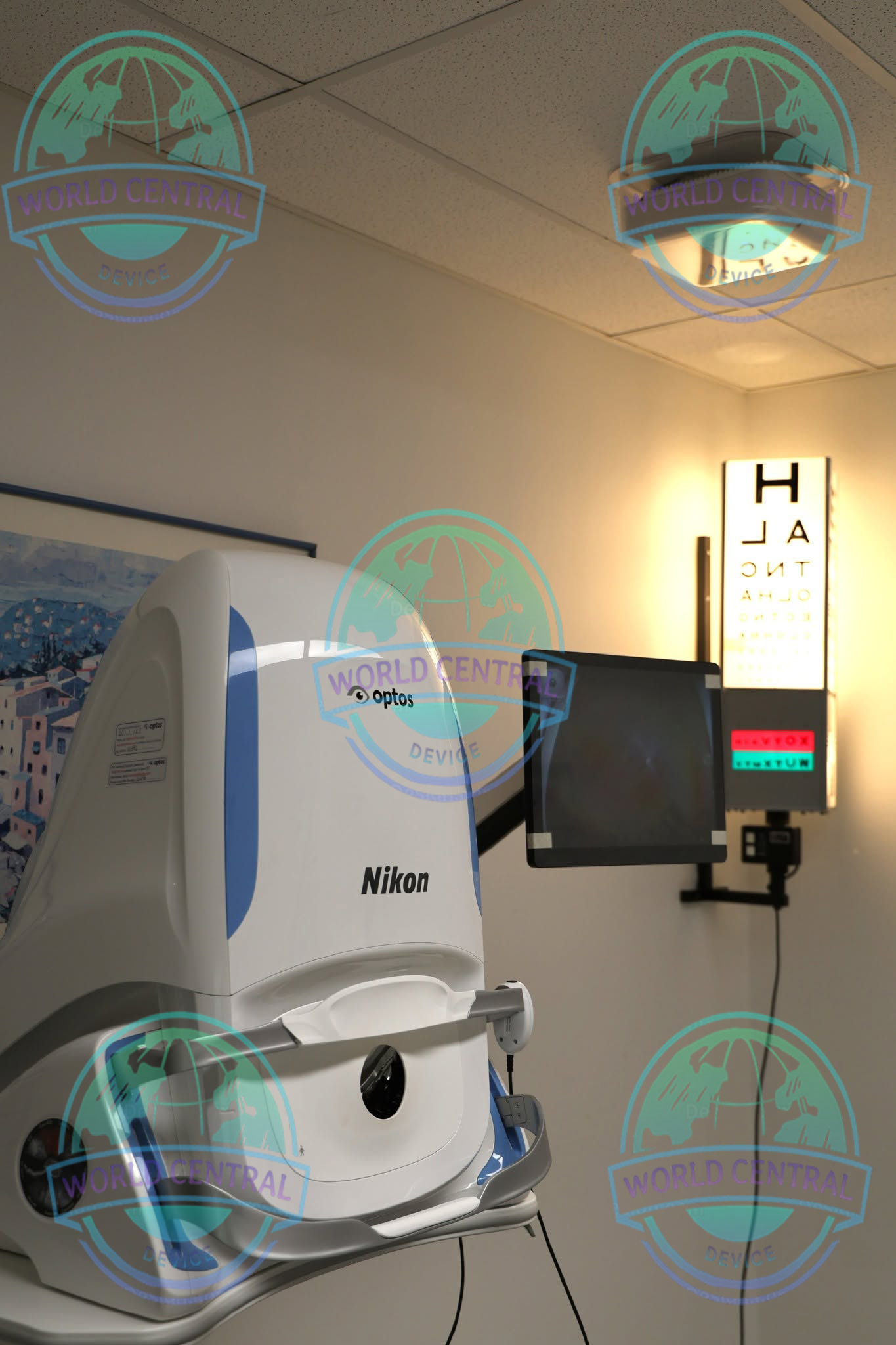
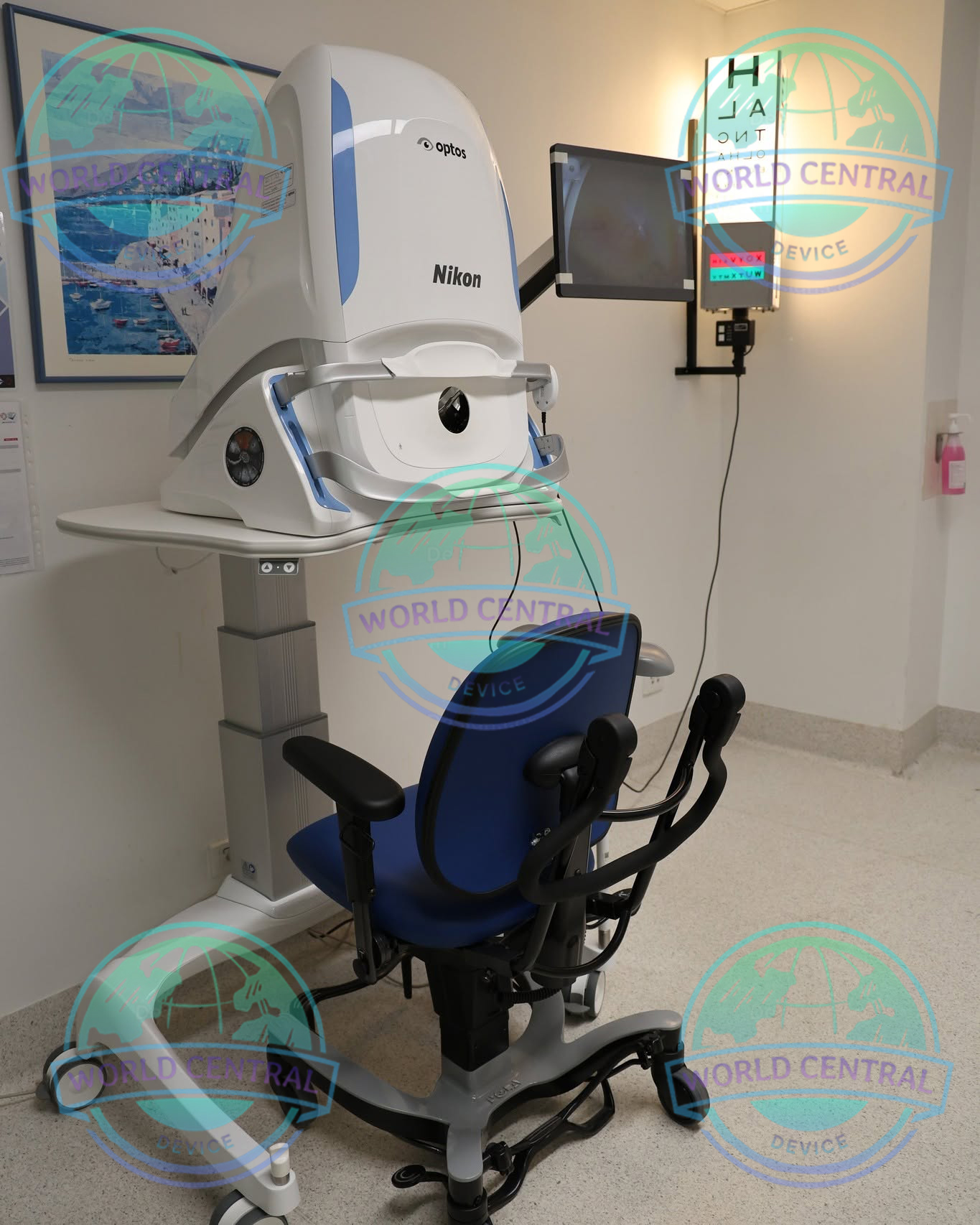
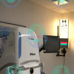
Optos Silverstone RGB Retinal Imaging
The Optos Silverstone RGB Retinal Imaging System is a premium multimodal ophthalmic imaging platform that combines ultra-widefield RGB retinal imaging with integrated Swept-Source Optical Coherence Tomography (SS-OCT)
Delivery and return
Delivery and return
Shipping Information
Shipping Information
Composition and care
Composition and care
Description
Optos Silverstone RGB Retinal Imaging System
Advanced Ultra-Widefield RGB Retinal Imaging with Integrated Swept-Source OCT
The Optos Silverstone RGB Retinal Imaging System is a premium multimodal ophthalmic imaging platform that combines true-color ultra-widefield retinal imaging with integrated Swept-Source Optical Coherence Tomography (SS-OCT). Developed to provide comprehensive retinal assessment in a single examination, the system enables eye care professionals to visualize both peripheral and central retinal pathology while obtaining deep, high-resolution cross-sectional retinal scans.
Designed for ophthalmologists, retina specialists, hospitals, academic institutions, and advanced eye care centers, the Silverstone RGB delivers exceptional image quality, efficient workflow, and advanced diagnostic capabilities. By integrating ultra-widefield optomap RGB imaging with swept-source OCT technology, clinicians can perform detailed retinal evaluations while improving disease detection, diagnosis, treatment planning, and long-term patient management.
Unlike conventional retinal imaging devices that primarily focus on the posterior pole, the Silverstone RGB captures an ultra-wide retinal view extending to the retinal periphery, allowing clinicians to identify pathology that might otherwise remain undetected.
Why Choose the Optos Silverstone RGB?
Modern retinal diagnostics require both comprehensive retinal visualization and detailed structural analysis.
The Optos Silverstone RGB combines these capabilities into one integrated platform, enabling clinicians to obtain ultra-widefield retinal images and high-resolution OCT scans during the same patient examination.
Key advantages include:
- Ultra-widefield RGB retinal imaging
- Integrated Swept-Source OCT
- Non-contact examination
- Non-mydriatic imaging capability for many patients
- High-speed image acquisition
- Deep retinal and choroidal visualization
- Simultaneous peripheral and macular assessment
- Advanced multimodal imaging
- Efficient digital workflow
- Comprehensive longitudinal patient monitoring
Key Features
Ultra-Widefield RGB Imaging
The Silverstone RGB utilizes advanced optomap® RGB imaging technology to capture high-resolution retinal images with realistic color representation.
The system provides visualization of approximately 200 degrees of the retina in a single capture, allowing clinicians to evaluate both central and peripheral retinal structures without the need for multiple image montages.
Clinical benefits include:
- Comprehensive retinal assessment
- Enhanced color fidelity
- Peripheral pathology detection
- Improved lesion documentation
- Better patient education
- Efficient disease monitoring
Image Modalities
optomap color rgb (red, green and blue laser)
optomap color rg (red and green laser)
optomap Sensory Retina (green laser)
optomap Choroidal (red laser)
optomap green af (green laser): autofluorescence
optomap blue af (blue laser): autofluorescence
optomap fa (blue laser): fluorescein angiography
optomap icg (infra-red): indocyanine green angiography
Swept Source Optical Coherence Tomography (OCT)
Resolution
optomap plus: 14 μm
optomap: 20 μm
Wavelengths
Red laser: 635 nm
Green laser: 532 nm (for af )
Blue laser: 488 nm (for fa)
Infra-red: 802 nm (for icg)
Integrated Swept-Source OCT
The integrated Swept-Source OCT (SS-OCT) provides high-speed cross-sectional retinal imaging with deeper tissue penetration than conventional spectral-domain OCT systems.
Swept-source technology enables detailed visualization of:
- Retinal layers
- Vitreoretinal interface
- Choroid
- Retinal pigment epithelium
- Optic nerve head
- Posterior vitreous
True Multimodal Imaging
The Silverstone RGB supports multiple imaging modalities that provide complementary diagnostic information during a single examination.
Available imaging modes include:
Ultra-Widefield RGB Color Imaging
Provides realistic retinal color representation with excellent visualization of retinal vessels, optic nerve, macula, and peripheral retina.
Applications include:
- Routine retinal examination
- Diabetic retinopathy
- Hypertensive retinopathy
- Peripheral retinal disease
- Retinal vascular disorders
Advanced Swept-Source OCT Technology
Swept-source OCT provides rapid acquisition speeds and improved penetration into deeper ocular tissues compared with many traditional OCT systems.
Key clinical capabilities include:
- High-resolution B-scans
- Macular cube scanning
- Dense raster scans
- Wide-field OCT localization
- Retinal thickness evaluation
- Structural retinal analysis
- Longitudinal follow-up registration
Clinical Applications
Diabetic Retinopathy
The combination of ultra-widefield imaging and SS-OCT supports comprehensive diabetic eye examinations by visualizing peripheral retinal lesions and assessing diabetic macular edema.
Clinical benefits include:
- Peripheral lesion detection
- Microaneurysm visualization
- Neovascularization assessment
- Macular edema evaluation
- Treatment planning
- Disease monitoring
Age-Related Macular Degeneration (AMD)
Silverstone RGB provides detailed structural and widefield imaging for evaluating both dry and neovascular AMD.
Applications include:
- Geographic atrophy monitoring
- Retinal pigment epithelium assessment
- Choroidal evaluation
- Longitudinal comparison
Glaucoma Management
Although glaucoma diagnosis requires multiple examinations, the Silverstone RGB contributes valuable structural documentation through retinal imaging and optic nerve visualization.
Applications include:
- Optic nerve documentation
- Retinal nerve fiber evaluation
- Digital progression analysis
- Clinical comparison
OCT Imaging
Signal Type: Optical scattering from tissue
Signal Source: Swept source OCT, Wavelength 1050 nm
Optical Power: Laser safety Class-1 following IEC/en60825-1:2014(2007)
Axial Resolution: <7 micron
Transverse Resolution: <20 micron (in tissue)
Scanners: Galavanometric with x, y pair
Scan Depth: Up to 2.5mm
General System Specifications
| Specification | Details |
|---|---|
| Product Name | Optos Silverstone RGB Retinal Imaging System |
| Brand | Optos (A Nikon Company) |
| Manufacturer | Optos plc |
| Product Family | Silverstone Series |
| Device Type | Ultra-Widefield Retinal Imaging System with Integrated Swept-Source OCT |
| Imaging Platform | Multimodal Digital Ophthalmic Imaging |
| Imaging Technology | optomap® Ultra-Widefield RGB Imaging + Swept-Source OCT |
| Optical Principle | Scanning Laser Ophthalmoscopy (SLO) |
| OCT Technology | Swept-Source Optical Coherence Tomography (SS-OCT) |
| Examination Type | Non-contact |
| Imaging Method | Single-Capture Ultra-Widefield Imaging |
| Clinical Application | Retinal Imaging & OCT Diagnostics |
| Intended Users | Ophthalmologists, Retina Specialists, Eye Hospitals |
| Patient Position | Seated |
| Operator Interface | Integrated Workstation |
| Patient Database | Included |
| Digital Workflow | Fully Integrated |
| Image Review | Real-Time |
| Image Comparison | Longitudinal Follow-up |
| Software Platform | Optos Proprietary Imaging Software |
| Image Storage | Digital |
| DICOM Support | Optional |
| EMR Connectivity | Supported |
| PACS Integration | Supported |
Ultra-Widefield RGB Imaging Specifications
| Parameter | Specification |
| Imaging Technology | optomap® RGB Imaging |
| Maximum Field of View | Up to approximately 200° |
| Retinal Coverage | Up to approximately 82% of retina |
| Capture Method | Single Capture |
| Image Capture Speed | Approximately 0.4 Seconds |
| Contact Imaging | No |
| Non-Mydriatic Imaging | Supported for many patients |
| Color Imaging | RGB True-Color Imaging |
| Peripheral Retina | Fully Visualized |
| Posterior Pole | Yes |
| Macula | Yes |
| Optic Disc | Yes |
| Retinal Vasculature | Yes |
| Peripheral Lesion Detection | Yes |
| Image Registration | Automatic |
| Image Enhancement | Digital |
| Image Magnification | Digital Zoom |
| Live Preview | Yes |
| Automatic Alignment | Yes |
| Internal Fixation | Yes |
| External Fixation | Optional |
Swept-Source OCT Specifications
| Parameter | Specification |
| OCT Technology | Swept-Source OCT |
| Scan Type | Cross-sectional Imaging |
| OCT Registration | Linked to optomap Image |
| Scan Localization | Ultra-Widefield Registered |
| Macular Cube Scan | Supported |
| Line Scan | Supported |
| High Density Raster Scan | Supported |
| Radial Scan | Supported |
| Retinal Thickness Evaluation | Yes |
| Retinal Layer Visualization | Yes |
| Choroid Visualization | Enhanced |
| Vitreoretinal Interface | Yes |
| Follow-up Registration | Automatic |
| Comparison Analysis | Yes |
| Image Averaging | Supported |
| Real-Time Scan Display | Yes |
| Structural Assessment | Yes |

There are no reviews yet.