
No products in the cart.: $0.00

On Sale!


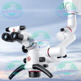
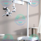
iSee 9000 Dental Microscope
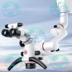
The iSee 9000 Dental Microscope is a professional surgical microscope engineered for modern dentistry. Featuring high-resolution apochromatic optics, powerful LED illumination, ergonomic positioning, and multiple magnification levels, it delivers exceptional visualization for endodontics, implantology, restorative dentistry, periodontics, prosthodontics
Delivery and return
Delivery and return
Shipping Information
Shipping Information
Composition and care
Composition and care
Description
iSee 9000 Dental Microscope
Professional Dental Surgical Microscope for Precision Dentistry
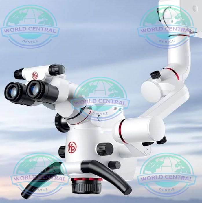
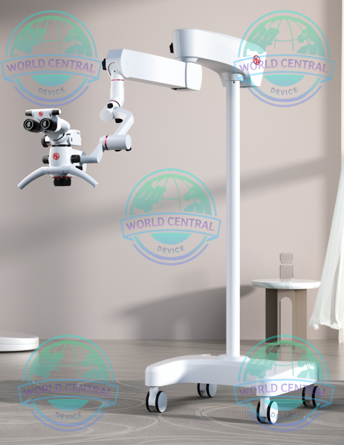
The iSee 9000 Dental Microscope is an advanced optical visualization system designed to enhance precision, ergonomics, and clinical confidence across a wide range of dental procedures. Engineered with high-definition apochromatic optics, powerful LED illumination, and a flexible ergonomic design, the microscope delivers exceptional image clarity while reducing operator fatigue during prolonged treatments.
Whether performing endodontic therapy, restorative dentistry, implant surgery, prosthodontics, or periodontal procedures, the iSee 9000 enables clinicians to visualize intricate anatomical structures that are often invisible to the naked eye. Its superior magnification and illumination improve diagnostic accuracy, treatment quality, and overall patient outcomes.
Modern dental practices increasingly rely on magnification technology to meet the growing demand for minimally invasive dentistry and predictable clinical results. The iSee 9000 combines premium optical performance with intuitive operation, making it suitable for private clinics, specialist practices, dental hospitals, universities, and teaching institutions.
Why Choose the iSee 9000 Dental Microscope?
Choosing a dental microscope is an investment in both clinical excellence and practitioner health. The iSee 9000 has been developed to provide outstanding visualization while supporting efficient workflows in everyday dental practice.
Its ergonomic construction helps maintain a natural working posture, minimizing neck, shoulder, and back strain commonly experienced during long procedures. Combined with crystal-clear optics and shadow-free illumination, clinicians can work with greater precision and confidence.
Key advantages include:
- High-definition apochromatic optical system
- Bright, uniform LED illumination
- Multiple magnification levels
- Superior depth of field
- Excellent color fidelity
- Ergonomic binocular viewing system
- Smooth multi-axis positioning
- Stable balancing arm mechanism
- Integrated documentation compatibility
- Designed for modern digital dentistry
Key Features
Imported SCHOTT Lens from Germany: Superior Optical Quality with SCHOTT lens + Apochromatic + Multi-layer optical coating design.
Crystal-clear display of every detail in the working area with better color accuracy and sensitivity to light.
Stepless Zoom System: Seamless view without interruptions.
Drum Type Magnification Changer: No interruption of the field of view during magnification adjustment.
Stepless/continuous magnification changer: Zoom factor from 0.4x to 2.4x for continuous zoom adjustment.
Uninterrupted field of view under the microscope for doctors.
Variable Working Distance: 270mm, covering the entire depth of the mouth without the need to move the microscope or change working position.
Quick and efficient focusing adjustment with only the objective lens button.
Imported Gas Shock Absorber from Germany: Super Balance Arm System for stability.
Integrated 4K Module in Video System: 4K High Resolution: 4096 x 2160 ultra-high resolution, providing images and videos with crystal clarity and lifelike colors.
Wireless foot pedal for quick and easy control.
Build Patient Trust: Some patients may be tense about receiving treatment.
Show them live images or playback supported by the integrated full HD camera and monitor to help them overcome the fear of the unknown.
Guiding patients throughout the general procedure can also be very helpful.
Record videos and images for each patient, allowing easy access to the complete medical history for every visit, strengthening patient trust further.
Flexible and Free Working: Pendulum design of the eyepiece tube maintains a horizontal observation position while the lens body can tilt left or right.
0-210° Inclined Eyepiece Tube: Adapt to various needs by achieving a comfortable posture in different operations.
Enhance ergonomics and work flexibility.
Effectively reduce and prevent muscle tension in the waist, neck, and shoulder areas.
Keep clinicians away from cervical spondylosis and lumbar spondylosis.
Premium Apochromatic Optical System
The microscope utilizes premium apochromatic optics that produce exceptionally sharp, distortion-free images with accurate color reproduction. Every anatomical detail can be observed with remarkable clarity, supporting more accurate diagnosis and treatment planning.
Benefits include:
- High optical resolution
- Minimal chromatic aberration
- Excellent contrast
- Natural color rendering
- Wide field of view
- Consistent image quality throughout the magnification range
Key benefits
Premium optics: German Schott apochromatic lenses with multilayer coating for superior color fidelity and detail.
Integrated 4K image: integrated 4K video module for real-time viewing, recording and sharing.
Continuous zoom: continuous magnification system from 0.4× to 19.6× without interruption of the field of view.
Varifocal objective lens: adjustable working distance up to 270 mm to cover the entire oral depth without repositioning the microscope.
Medical LED lighting: long lifespan (up to 60,000 hours), high color rendering index, and reduced eye strain.
Dedicated filters: green filter for visualizing vessels and nerves; orange filter to prevent premature curing of composites.
Advanced ergonomics: gas spring arm with cushioning for perfect balance; 0–210° tilting binocular tube and pendulum mode for comfortable posture.
Digital workflow: 4K recording to SD card/USB, easy case sharing, and team collaboration.
Multi-Step Magnification System
The iSee 9000 offers multiple magnification levels that allow clinicians to seamlessly transition from general observation to highly detailed microsurgical procedures.
Lower magnifications provide an excellent overview of the operative field, while higher magnifications enable visualization of microscopic structures such as:
- Root canal anatomy
- Microfractures
- Accessory canals
- Marginal restorations
- Crown preparation details
- Surgical incision margins
- Fine sutures
High-Intensity LED Illumination
Powerful coaxial LED illumination ensures bright, shadow-free lighting directly along the optical axis.
Compared with traditional halogen systems, LED illumination offers several advantages:
- Longer service life
- Lower heat generation
- Stable color temperature
- Reduced maintenance
- Instant brightness
- Consistent illumination intensity
- Improved patient comfort
Clinical Applications
Endodontics
Microscope-assisted endodontics has become the standard for advanced root canal therapy.
The iSee 9000 assists clinicians in:
- Locating calcified canals
- Detecting accessory canals
- Identifying vertical root fractures
- Removing separated instruments
- Performing apical microsurgery
- Evaluating obturation quality
- Managing complex retreatment cases
Restorative Dentistry
High magnification enables precise restorative procedures including:
- Margin evaluation
- Caries detection
- Composite layering
- Ceramic restoration placement
- Crown preparation
- Inlay and onlay fitting
- Adhesive procedures
Implant Dentistry
Implant surgeons benefit from enhanced visualization throughout the surgical workflow.
Applications include:
- Implant site preparation
- Soft tissue management
- Bone grafting
- Sinus lift procedures
- Guided implant surgery
- Implant uncovering
- Peri-implant tissue evaluation
Periodontal Surgery
The iSee 9000 supports delicate periodontal procedures by providing superior visualization of soft tissue structures.
Typical procedures include:
- Gingival grafting
- Flap surgery
- Connective tissue grafts
- Crown lengthening
- Periodontal regeneration
- Microsuturing
Prosthodontics
Microscopic visualization improves prosthetic precision throughout restorative treatment.
Applications include:
- Veneer preparation
- Crown preparation
- Bridge evaluation
- Impression verification
- Cement removal
- Occlusal assessment
- Margin finishing
Key specifications
Video resolution: 4K (4096×2160)
Zoom range: 0.4× to 19.6× (continuous and step-by-step)
Objective lens: varifocal, working distance up to 270 mm
Lighting: Medical LED, high CRI, lifespan up to 60,000 hours.
Control: Wireless foot pedal for recording and capturing.
Storage: SD / high-speed U disk
Optional accessories: extender, splitter/beam splitter, digital camera adapter, mobile phone adapter, “four-in-one” module.


There are no reviews yet.