
No products in the cart.: $0.00

On Sale!


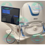
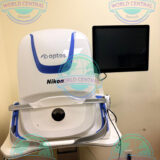
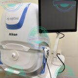
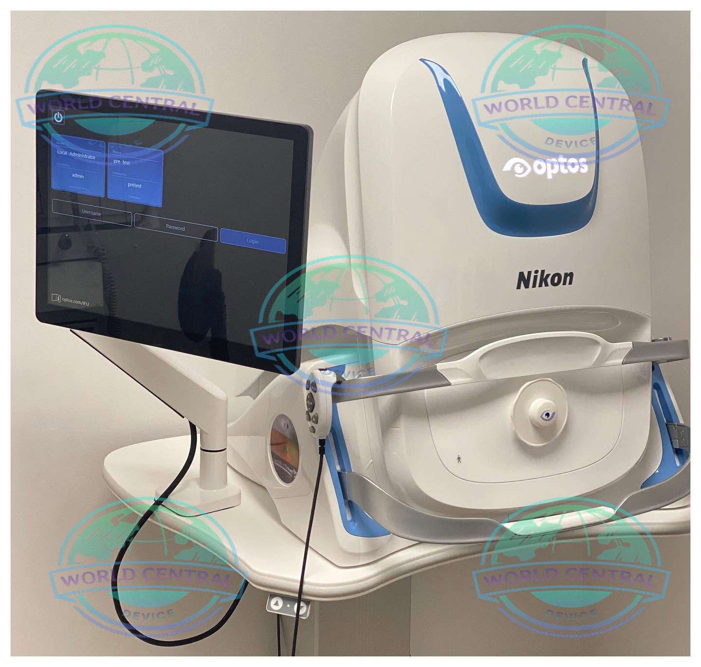
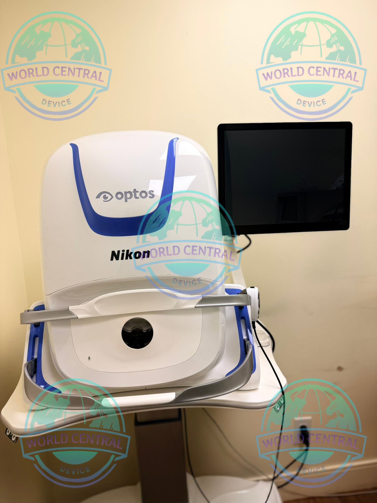
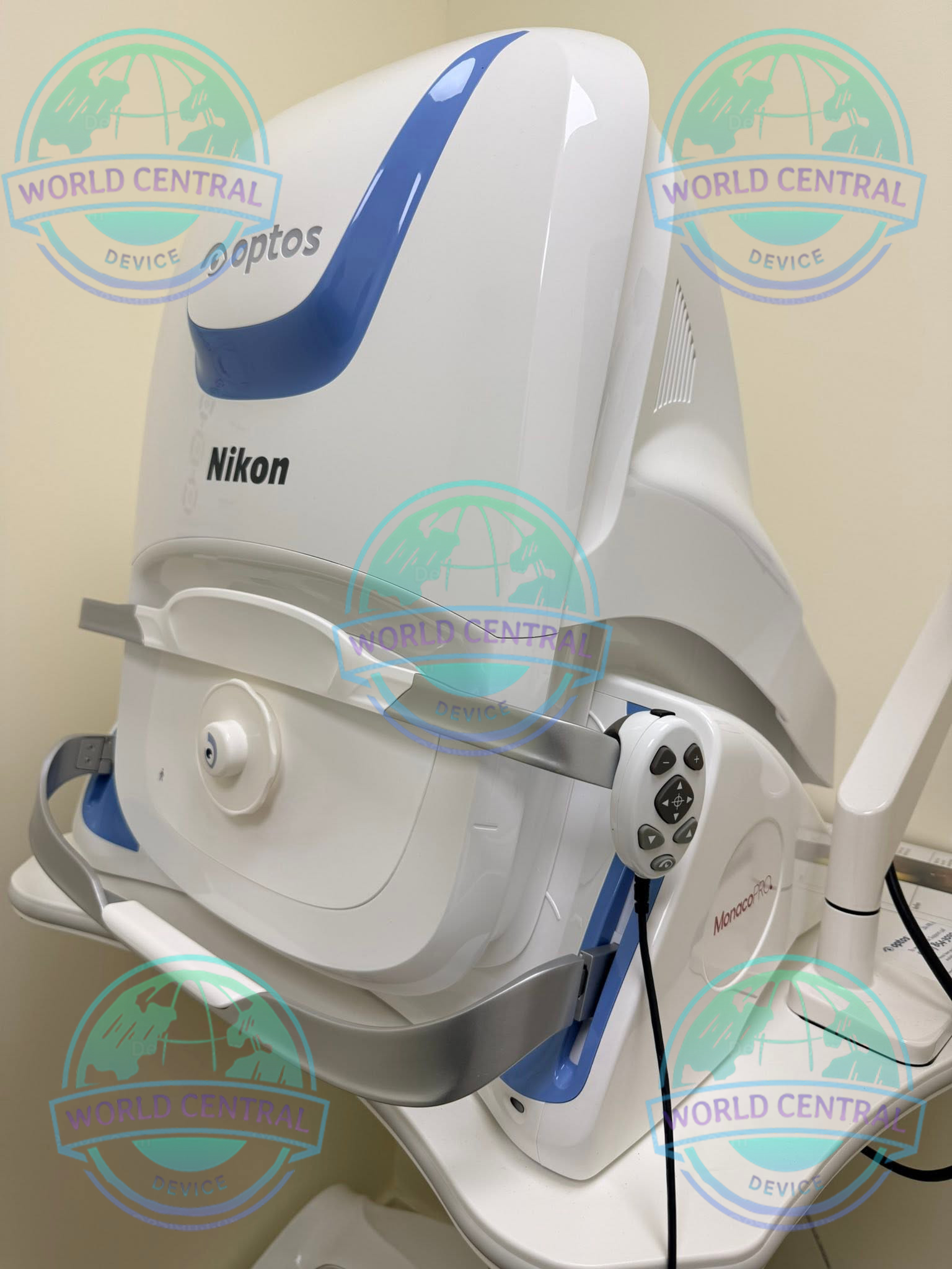
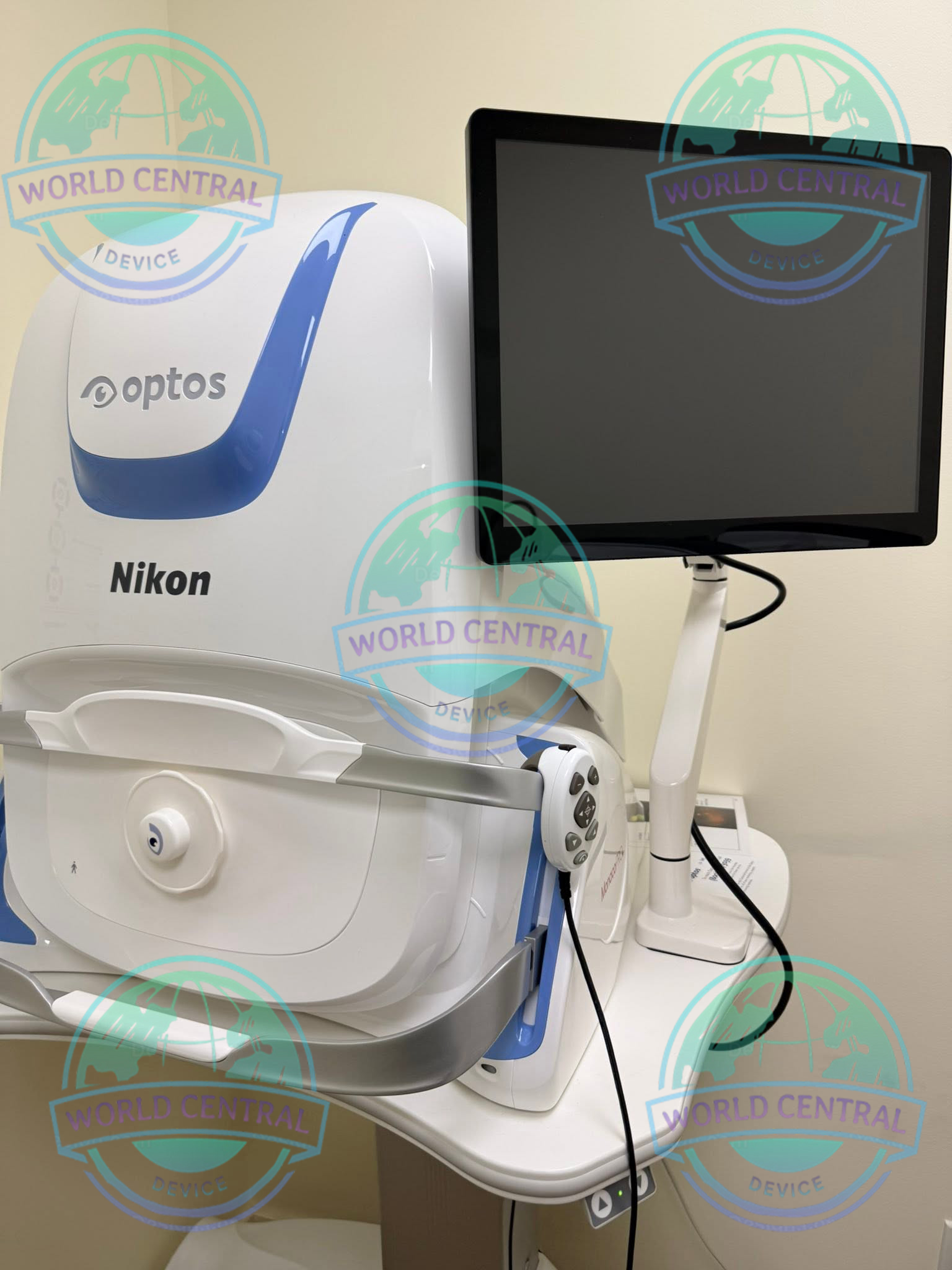
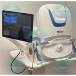
Nikon Optos Monaco Ultra-Widefield Retinal Imaging System
The Nikon Optos Monaco Ultra-Widefield Retinal Imaging System is an advanced ophthalmic imaging solution that combines ultra-widefield retinal imaging with integrated Spectral-Domain Optical Coherence Tomography (SD-OCT).
Delivery and return
Delivery and return
Shipping Information
Shipping Information
Composition and care
Composition and care
Description
Nikon Optos Monaco Ultra-Widefield Retinal Imaging System
Advanced Ultra-Widefield Retinal Imaging with Integrated Spectral-Domain OCT
The Nikon Optos Monaco Ultra-Widefield Retinal Imaging System is an advanced multimodal ophthalmic imaging platform that combines ultra-widefield retinal imaging with high-resolution Spectral-Domain Optical Coherence Tomography (SD-OCT). Designed for comprehensive retinal examination and efficient clinical workflow, Monaco enables eye care professionals to visualize both peripheral and central retinal pathology within a single integrated system.
By combining Optos’ patented ultra-widefield (UWF) optomap® technology with precise SD-OCT cross-sectional imaging, the Monaco system supports earlier disease detection, improved diagnosis, and more effective long-term monitoring of retinal conditions. The platform captures detailed retinal images without requiring extensive pupil dilation in many patients, improving both patient comfort and clinical efficiency.
Ideal for ophthalmology clinics, retina specialists, hospitals, research institutions, and academic medical centers, the Nikon Optos Monaco delivers exceptional imaging performance across a broad spectrum of retinal diseases, including diabetic retinopathy, glaucoma, retinal vascular disorders, macular degeneration, peripheral retinal lesions, and inherited retinal diseases.
Why Choose the Nikon Optos Monaco?
Modern retinal care requires more than traditional fundus photography. Clinicians need comprehensive visualization, advanced diagnostic tools, and streamlined workflow to accurately evaluate both peripheral and central retinal pathology.
The Nikon Optos Monaco provides an all-in-one imaging solution by integrating multiple imaging technologies into a single workstation.
Key advantages include:
- Ultra-widefield retinal imaging up to approximately 200°
- Integrated Spectral-Domain OCT
- Non-contact image acquisition
- Non-mydriatic imaging capability for many patients
- High-speed OCT scanning
- Multimodal retinal imaging
- Simultaneous central and peripheral retinal assessment
- Excellent patient comfort
- Efficient clinical workflow
- Digital image management and follow-up comparison
Features and Benefits
UWF with integrated OCT saves time, space and minimizes patient movement.
High resolution 200º single-capture optomap images improve pathology detection and management frommacula through the far periphery.
Non-mydriatic, cSlO imaging through most cataracts and small pupils (2 mm).
3-in-1 Color Depth ImagingTM provides important clinical data from the retinal surface through the choroid.
Green laser autofluorescence minimizes patient exposure to blue light and shows macula and optic nerve head detail.
Central pole OCT provides comprehensive multi-modal imaging.
optomap images and OCT scans are correlated to facilitate pathology examination.
Color, AF, and OCT images are shown in a single, comprehensive view.
Fast, comfortable image acquisition is easier on patients and improves clinic flow.
OptosAdvance Image Management facilitates image review and consultations and includes measurement and auto montage capabilities.
Image Modalities
-Color
-Red-free
-Choroidal
-Autofluorescence
-OCT
Image Views
-Standard: 200⁰ Single Capture
-Auto-montage: Up to 220⁰
-Central Pole: Detailed view of the macula
-Stereo: Image pairing for optic disc and retinal evaluation
-OCT: Cross-sectional imaging of ocular structures, including the fundus
Integrated Spectral-Domain OCT
Monaco combines ultra-widefield imaging with advanced Spectral-Domain Optical Coherence Tomography (SD-OCT), allowing clinicians to evaluate retinal microstructures with exceptional detail.
High-resolution OCT imaging provides cross-sectional visualization of retinal layers, enabling early identification of subtle pathological changes.
Clinical applications include:
- Macular edema evaluation
- Age-related macular degeneration assessment
- Vitreomacular interface disorders
- Epiretinal membrane analysis
- Macular hole diagnosis
- Central serous chorioretinopathy
- Glaucoma structural assessment
Tomographic Imaging
Signal Type: Optical scattering from tissue
Signal Source: Super luminescent Diode (SlD) 830 nm
Optical Power: laser safety Class-1 following IEC/en60825-1:2014
Typical Axial Resolution: <10 micron (in tissue) Digital on-screen <6 micron
Transverse Resolution: 20 micron (in tissue)
Scanners: Galavanometric with x, y mirrors
Scan Depth: Up to 2.5mm
OCT Scan Characteristics
Spectral Domain OCT
A-Scan rate up to 70k cycles/s
Active eye tracking
Automatic scan positioning
OCT Scan Types
Line Scan
Raster Scan
Retina Topography Scan
Optic nerve Head (OnH) Topography Scan
Retinal nerve Fiber layer (RnFl) Scan
Footprint
Width: 550 mm/22 inches
Depth: 500 mm/19.5 inches
Height: 650 mm/25.5 inches
Technical Specifications
Imaging System Specifications
| Specification | Details |
|---|---|
| Product Name | Nikon Optos Monaco Ultra-Widefield Retinal Imaging System |
| Imaging Technology | Ultra-Widefield Scanning Laser Ophthalmoscopy (UWF SLO) + Spectral-Domain Optical Coherence Tomography (SD-OCT) |
| Imaging Type | Multimodal Retinal Imaging |
| Fundus Imaging | Ultra-Widefield Digital Imaging |
| OCT Technology | Spectral-Domain OCT |
| Image Acquisition | Non-contact |
| Patient Position | Seated |
| Alignment | Motor-assisted alignment with live fundus view |
| Image Capture | Single-shot retinal imaging |
| Clinical Use | Diagnostic retinal imaging and follow-up |
| Image Registration | Integrated UWF image with OCT scan localization |
| Digital Workflow | Yes |
| DICOM Support | Available (configuration dependent) |
| Electronic Medical Record Integration | Supported (configuration dependent) |
Ultra-Widefield Imaging Specifications
| Parameter | Specification |
| Maximum Retinal Capture | Up to approximately 200° in a single capture |
| Retinal Coverage | Up to approximately 82% of the retina in one image |
| Image Acquisition Time | Approximately 0.4 second |
| Pupil Dilation | Often not required (depends on patient and clinical conditions) |
| Peripheral Retina Visualization | Yes |
| Macular Visualization | Yes |
| Optic Disc Visualization | Yes |
| Retinal Periphery Imaging | Yes |
| Image Magnification | Digital |
| Image Export | JPEG, TIFF, DICOM (configuration dependent) |
Spectral-Domain OCT Specifications
| Parameter | Specification |
| OCT Type | Spectral-Domain Optical Coherence Tomography |
| Scan Mode | Cross-sectional imaging |
| Macular Cube Scan | Supported |
| Line Scan | Supported |
| High-Density Raster Scan | Supported |
| B-Scan Imaging | Supported |
| Retinal Thickness Analysis | Supported |
| Retinal Layer Visualization | Supported |
| Follow-up Scan Registration | Supported |
| Image Averaging | Supported |
| OCT Localization | Registered to ultra-widefield image |
Laser Imaging Technology
| Parameter | Specification |
| Imaging Principle | Scanning Laser Ophthalmoscopy |
| Green Laser | Used for visualization of superficial retinal structures |
| Red Laser | Used for deeper retinal and choroidal visualization |
| Composite Image Generation | Yes |
| Laser Safety | Medical ophthalmic laser system compliant with applicable standards |
| Confocal Imaging | Yes |
| Optical Alignment | Automatic assistance |
Multimodal Imaging Modes
| Imaging Mode | Availability |
| Ultra-Widefield Color Imaging | Yes |
| Red Laser Imaging | Yes |
| Green Laser Imaging | Yes |
| Pseudocolor Imaging | Yes |
| Fundus Autofluorescence (FAF) | Yes |
| SD-OCT | Yes |
| Image Overlay | Yes |
| Comparative Follow-up | Yes |
| Side-by-Side Image Review | Yes |
| Historical Image Comparison | Yes |
Optical Specifications
| Parameter | Specification |
| Imaging Optics | Scanning Laser Optical System |
| Focusing | Automatic with manual adjustment capability |
| Fixation Targets | Internal fixation with selectable targets |
| Patient Eye | Right and Left Eye |
| Image Resolution | High-resolution digital imaging |
| Optical Path | Integrated retinal imaging and OCT |
| Internal Calibration | Factory calibrated |

There are no reviews yet.