
No products in the cart.: $0.00

On Sale!


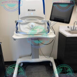
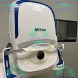
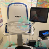
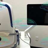
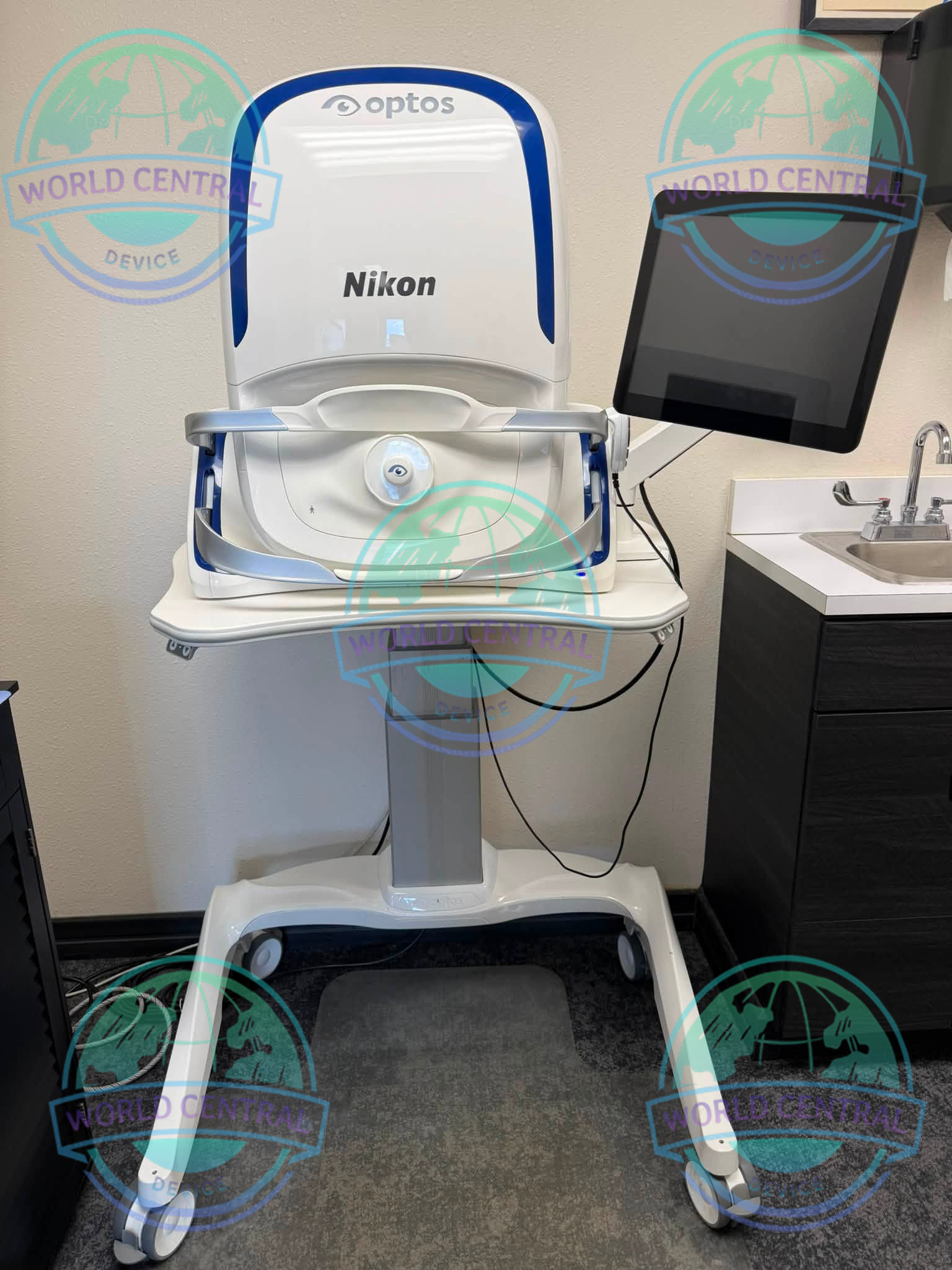
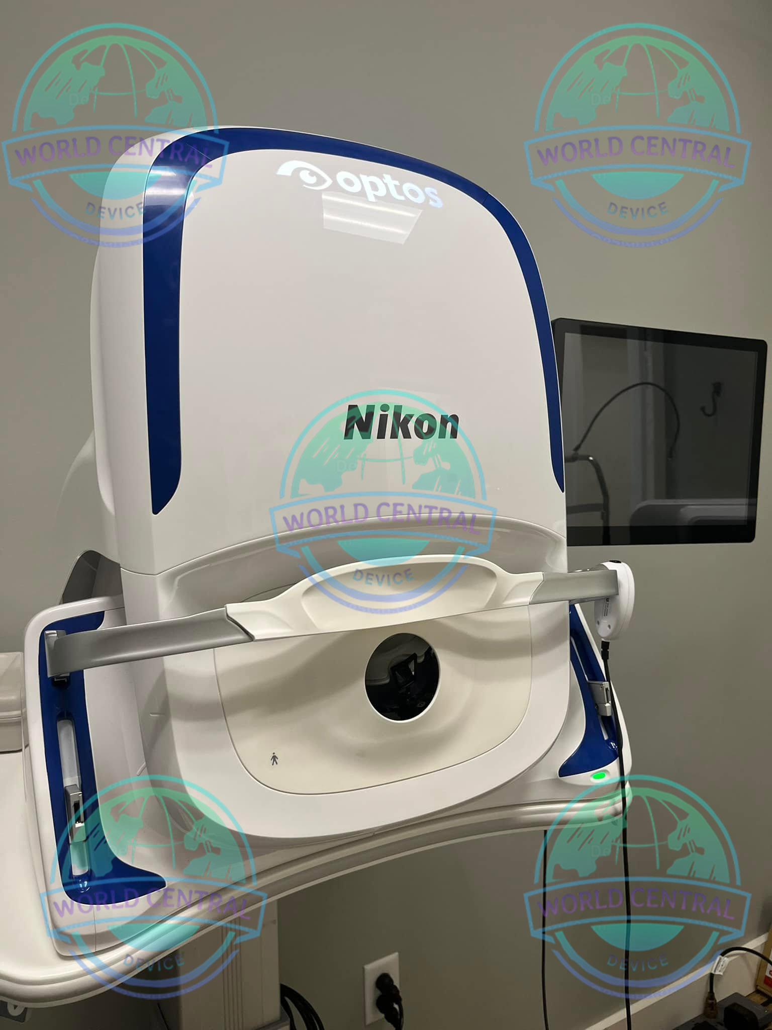
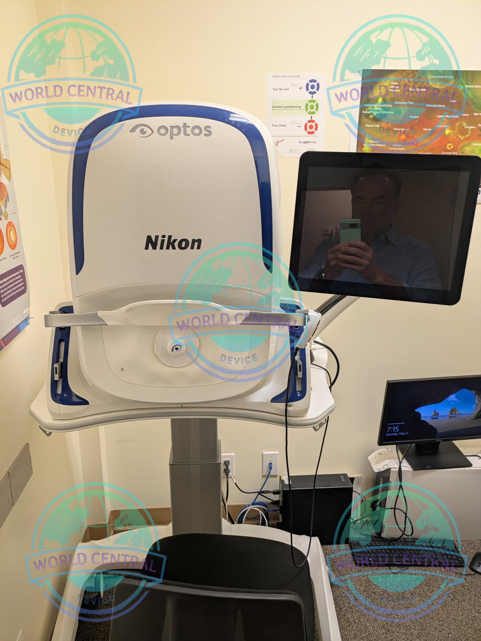
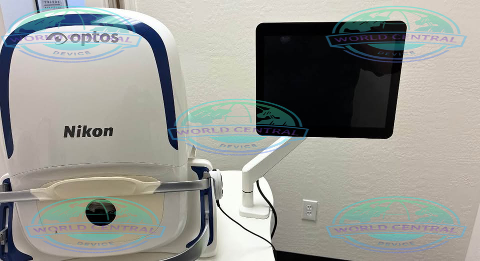
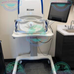
Optos California Retinal Imaging System
The Optos California Retinal Imaging System is an advanced ultra-widefield (UWF) retinal imaging solution that delivers fast, high-resolution visualization of up to approximately 200° of the retina in a single capture
Delivery and return
Delivery and return
Shipping Information
Shipping Information
Composition and care
Composition and care
Description
Optos California Retinal Imaging System
Advanced Ultra-Widefield Retinal Imaging for Comprehensive Ophthalmic Diagnostics
The Optos California Retinal Imaging System is a premium ultra-widefield (UWF) retinal imaging platform engineered to deliver fast, high-resolution visualization of the retina with exceptional clinical efficiency. Utilizing Optos scanning laser ophthalmoscopy technology, the system captures up to approximately 200 degrees of the retina in a single image, enabling eye care professionals to assess both the central and peripheral retina during one examination.
Designed for modern ophthalmology practices, retina specialists, hospitals, and academic institutions, the Optos California combines ultra-widefield imaging with multimodal diagnostic capabilities, including color imaging, fundus autofluorescence (FAF), red-free imaging, and choroidal imaging. These imaging modalities help clinicians detect, diagnose, document, and monitor a wide range of retinal diseases with greater confidence.
Its non-contact, patient-friendly design minimizes examination time while improving workflow efficiency. In many patients, high-quality retinal images can be obtained without pharmacologic pupil dilation, making the examination more comfortable and convenient.
Why Choose the Optos California Retinal Imaging System?
Retinal disorders often develop beyond the posterior pole, making conventional fundus photography insufficient for comprehensive retinal evaluation. The Optos California expands retinal visualization by capturing a significantly larger retinal field in a single acquisition, supporting earlier disease detection and more accurate long-term monitoring.
Key advantages include:
- Ultra-widefield retinal imaging up to approximately 200°
- Visualization of central and peripheral retina in one image
- Non-contact imaging technology
- Non-mydriatic imaging capability for many patients
- Multiple imaging modalities in one platform
- High-resolution digital retinal imaging
- Fast image acquisition
- Comfortable patient experience
- Efficient clinical workflow
- Advanced image management software
- Longitudinal patient follow-up
Key Features
Ultra-Widefield Retinal Imaging
The Optos California employs advanced scanning laser ophthalmoscopy (SLO) technology to obtain ultra-widefield retinal images with exceptional clarity.
Unlike conventional fundus cameras that typically capture only a limited central retinal field, the Optos California enables clinicians to examine both posterior and peripheral retinal structures in a single capture.
Clinical benefits include:
- Expanded retinal visualization
- Earlier peripheral pathology detection
- Improved retinal documentation
- Better disease monitoring
- Reduced need for image montages
- Enhanced clinical confidence
What it Can Detect
Optomap Images, using the OPTOS Californa, allow doctors to see a full view of the retina and apply different light filters to view various layers of the retina. This allows for quick and more accurate diagnosis of retinal disorders compared to traditional methods. Common eye diseases that can bee detected using the OTPOS California include:
Retinal Disease
Macular degeneration (build-up called drusen in the central vision)
Glaucoma (damage to the optic nerve)
Diabetic retinopathy (leaking or bleeding inside the eyes)
Hypertensive retinopathy (constricted blood vessels or bleeding)
Nevi (freckles)
Melanoma (cancers)
Retinal Condition
Holes
Tears
Retinal degenerations (genetic or age related)
Retinal thinning (lattice)
Retinal Detachment
Image Modalities
Color rg
Color rgb (available with rg/af/fa combined model)
Sensory Red-free
Choroidal
Autofluorescence (green and blue*)(*feature may not be available in all regions)
Fluorescein angiography
Indocyanine green angiography
Specifications
Image Modalities: optomap color and optomap plus (red and green laser):
Color composite view
Green laser view
Red laser view
optomap af (green laser): autofluorescence
Resolution: optomap color: 20 μm, optomap plus, af : 14 μm
Wavelengths: Red laser: 635 nm, Green laser: 532 nm
Exposure Time: Less than 0.4 seconds
Foot Print: Width: 550 mm / 22 inches
Depth: 550 mm / 22 inches including chinrest
Height: 608-632 mm / 24-25 inches
Weight: 75 lbs
Communication Protocol: DICOM Compatible
General System Specifications
| Specification | Details |
|---|---|
| Product Name | Optos California Retinal Imaging System |
| Brand | Optos (A Nikon Company) |
| Manufacturer | Optos plc |
| Device Type | Ultra-Widefield Retinal Imaging System |
| Imaging Technology | Scanning Laser Ophthalmoscopy (SLO) |
| Imaging Platform | Digital Multimodal Retinal Imaging |
| Examination Type | Non-contact Retinal Imaging |
| Clinical Application | Ophthalmic Diagnostic Imaging |
| Intended Users | Ophthalmologists, Retina Specialists, Eye Care Professionals |
| Installation | Floor-standing Clinical Imaging System |
Ultra-Widefield Imaging Specifications
| Parameter | Specification |
| Maximum Retinal Field of View | Up to approximately 200° |
| Retinal Coverage | Up to approximately 82% of the retina in one image |
| Image Acquisition | Single Capture |
| Capture Speed | Approximately 0.4 seconds |
| Peripheral Retina Visualization | Yes |
| Central Retina Imaging | Yes |
| Macular Imaging | Yes |
| Optic Disc Imaging | Yes |
| Retinal Periphery Assessment | Yes |
| Non-Mydriatic Imaging | Supported for many patients |
| Contact Lens Required | No |
| Patient Comfort | High |
Imaging Modalities
| Imaging Mode | Availability |
| Ultra-Widefield Color Imaging | Yes |
| optomap® Imaging | Yes |
| Fundus Autofluorescence (FAF) | Yes |
| Red-Free Imaging | Yes |
| Choroidal Imaging | Yes |
| Digital Color Composite | Yes |
| Image Overlay | Yes |
| Comparative Review | Yes |
| Historical Image Comparison | Yes |
Optical System Specifications
| Parameter | Specification |
| Optical Principle | Scanning Laser Ophthalmoscopy |
| Optical Alignment | Automatic |
| Internal Fixation Target | Yes |
| External Fixation | Optional |
| Focus Adjustment | Automatic with Manual Override |
| Optical Calibration | Factory Calibrated |
| Image Processing | Digital |
| Eye Selection | Right / Left Eye |
Laser Imaging Specifications
| Parameter | Specification |
| Imaging Method | Dual-Wavelength Scanning Laser |
| Green Laser | Superficial retinal visualization |
| Red Laser | Deep retinal & choroidal visualization |
| Composite Image Generation | Yes |
| Laser Classification | Medical Ophthalmic Laser |
| Laser Safety | IEC-compliant (market dependent) |
Clinical Imaging Capabilities
| Clinical Assessment | Supported |
| Diabetic Retinopathy | ✔ |
| Age-Related Macular Degeneration | ✔ |
| Glaucoma Documentation | ✔ |
| Retinal Vein Occlusion | ✔ |
| Retinal Artery Occlusion | ✔ |
| Peripheral Retinal Lesions | ✔ |
| Retinal Tears | ✔ |
| Retinal Detachment Assessment | ✔ |
| Choroidal Lesions | ✔ |
| Ocular Oncology Documentation | ✔ |
| Uveitis Documentation | ✔ |
| Retinal Dystrophies | ✔ |

There are no reviews yet.