
No products in the cart.: $0.00

On Sale!

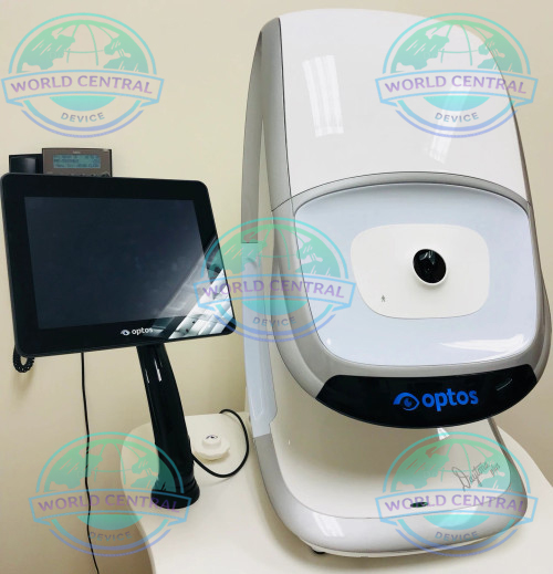
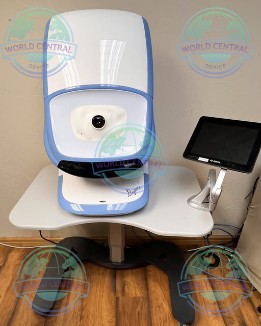
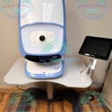
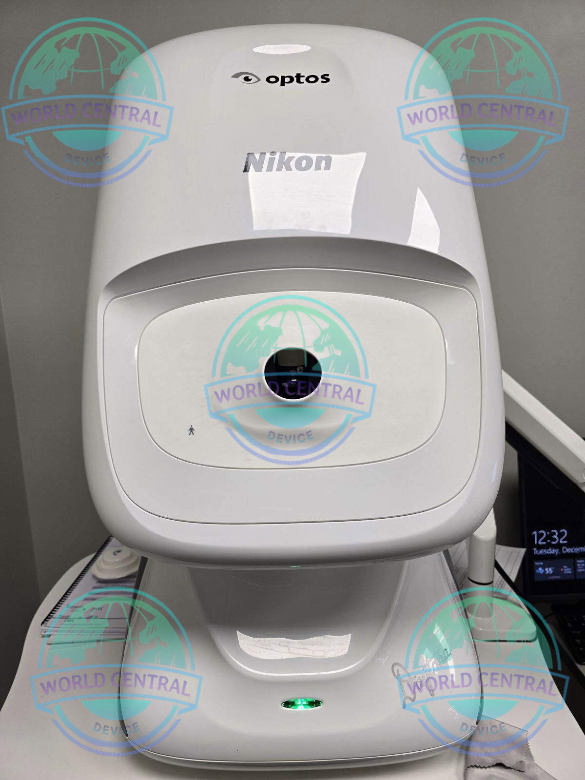
Optos Daytona Retinal Imaging
The Optos Daytona Retinal Imaging System is an advanced ultra-widefield retinal imaging solution designed for comprehensive ophthalmic diagnostics. Utilizing Optos’ patented optomap scanning laser ophthalmoscopy (SLO) technology
Delivery and return
Delivery and return
Shipping Information
Shipping Information
Composition and care
Composition and care
Description
Optos Daytona Retinal Imaging System
Ultra-Widefield Retinal Imaging for Fast, Comprehensive Eye Examinations
The Optos Daytona Retinal Imaging System is an advanced ultra-widefield (UWF) retinal imaging device designed to provide high-resolution visualization of the retina in a fast, patient-friendly, and non-contact examination. Powered by Optos’ patented scanning laser ophthalmoscopy (SLO) technology, the Daytona captures up to approximately 200 degrees of the retina in a single image, allowing clinicians to evaluate both the central and peripheral retina without the need for multiple image montages.
Developed for ophthalmologists, retina specialists, optometrists, eye hospitals, and vision care centers, the Optos Daytona enhances diagnostic confidence by providing comprehensive retinal imaging in just seconds. The system supports routine eye examinations, disease screening, clinical documentation, and long-term monitoring of retinal conditions.
With its compact footprint, intuitive workflow, and non-mydriatic imaging capability for many patients, the Optos Daytona is an ideal solution for busy clinical environments seeking efficient, high-quality retinal imaging.
Why Choose the Optos Daytona Retinal Imaging System?
Traditional fundus cameras often provide a limited view of the retina, requiring multiple images to assess peripheral pathology. The Optos Daytona overcomes this limitation by delivering an ultra-widefield retinal image that captures a significantly larger retinal area in a single acquisition.
Key benefits include:
- Ultra-widefield retinal imaging up to approximately 200°
- Visualization of approximately 82% of the retina in one image
- Non-contact examination
- Non-mydriatic imaging capability for many patients
- High-speed image acquisition
- Compact and space-saving design
- Excellent patient comfort
- Digital image management
- Efficient clinical workflow
- Reliable longitudinal patient follow-up
Key Features
-200° ultra-widefield imaging in less than 0.4 seconds
-Non-mydriatic and non-contact imaging through 2 mm pupils
-High-resolution optomap images (14–20 µm)
-Autofluorescence with green laser to show lipofuscin in RPE
-3-in-1 Color Depth imaging: Color, Sensory (red-free), and Choroidal
-DICOM compatible and software with advanced visualization tools
-Includes stereo disc imaging and 3D wrap for patient education
-Images available immediately and suitable for telehealth
Ultra-Widefield Retinal Imaging
The Optos Daytona utilizes advanced scanning laser ophthalmoscopy (SLO) technology to capture detailed retinal images extending far into the retinal periphery.
Unlike conventional fundus photography, the Daytona enables clinicians to visualize peripheral retinal structures that are often associated with early retinal pathology.
Clinical advantages include:
- Expanded retinal visualization
- Earlier detection of peripheral abnormalities
- Improved documentation
- Better disease monitoring
- Enhanced diagnostic confidence
- Reduced need for image stitching
Non-Contact Imaging Technology
Patient comfort is a major advantage of the Optos Daytona.
The non-contact imaging design eliminates direct eye contact with the imaging system, improving patient experience while reducing examination complexity.
Benefits include:
- Comfortable examination
- Reduced patient anxiety
- Faster patient positioning
- Improved workflow efficiency
- Simplified image acquisition
Non-Mydriatic Imaging
For many patients, high-quality retinal images can be obtained without pharmacologic pupil dilation.
Advantages include:
- Shorter examination time
- Greater patient convenience
- Improved clinic throughput
- Reduced waiting time
- Enhanced patient satisfaction
High-Resolution Digital Imaging
The Daytona produces high-quality digital retinal images suitable for diagnosis, documentation, patient education, referrals, and follow-up examinations.
The imaging system delivers excellent visualization of:
- Macula
- Optic nerve head
- Retinal vessels
- Peripheral retina
- Choroidal structures
- Retinal pigment epithelium
optomap Imaging Technology
Optos’ proprietary optomap® technology enables clinicians to acquire a comprehensive retinal image in approximately 0.4 seconds.
The technology supports:
- Rapid image acquisition
- High patient throughput
- Consistent image quality
- Efficient clinical documentation
- Longitudinal image comparison
Clinical Applications
Diabetic Retinopathy
The Optos Daytona supports comprehensive diabetic eye examinations by visualizing both central and peripheral retinal lesions.
Applications include:
- Diabetic retinopathy screening
- Peripheral lesion detection
- Hemorrhage documentation
- Microaneurysm assessment
- Disease progression monitoring
- Referral support
| Feature | Specification |
|---|---|
| Condition | Pre-owned (Reconditioned) |
| Model | Optos Daytona P200T |
| Image Capture | 200° ultra-widefield (82% retina) |
| Imaging Modalities | Color, Sensory (red-free), Choroidal, Autofluorescence |
| Resolution | Optomap: 20 µm, Optomap Plus: 14 µm |
| Wavelengths | Red laser: 635 nm, Green laser: 532 nm |
| Exposure Time | Less than 0.4 seconds |
| Display | Screen and tablet included |
| Software | 3-in-1 Color Depth, Autofluorescence, EyeSteering, 3D Wrap |
| Communication | DICOM compatible |
| Dimensions | 440 mm (W) x 500 mm (D) x 795 mm (H) |
| Weight | 28 kg (62 lbs) |
| Table Dimensions | 887 mm (W) x 600 mm (D) |
| System Voltage | US: 100–120V / EU: 200–240V |
| Power Consumption | 300 VA |
| Laser Safety | Class 1 (EN60825-1, 21 CFR 1040.10/.11) |
| Included Accessories | Motorized table, computer, screen, tablet |
| Warranty | 6 months |
| Availability | Pick-up only |


There are no reviews yet.